Movie
Movie Controller


+ Open data

- Basic information
| Entry |  | |||||||||
|---|---|---|---|---|---|---|---|---|---|---|
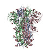
| Title | Sarbecovirus HeB2013 Spike Trimer in a Locked Conformation | |||||||||
 Map data Map data | ||||||||||
 Sample Sample |
| |||||||||
 Keywords Keywords | spike protein / viral protein | |||||||||
| Function / homology |  Function and homology information Function and homology informationhost cell endoplasmic reticulum-Golgi intermediate compartment membrane / receptor-mediated virion attachment to host cell / endocytosis involved in viral entry into host cell / fusion of virus membrane with host plasma membrane / fusion of virus membrane with host endosome membrane / viral envelope / host cell plasma membrane / virion membrane / membrane Similarity search - Function | |||||||||
| Biological species |  BtRf-BetaCoV/HeB2013 (virus) BtRf-BetaCoV/HeB2013 (virus) | |||||||||
| Method | single particle reconstruction / cryo EM / Resolution: 3.45 Å | |||||||||
 Authors Authors | Wang J / Xiong X | |||||||||
| Funding support | 2 items
| |||||||||
 Citation Citation | Journal: Sci Adv / Year: 2025 Title: SARS-related coronavirus S-protein structures reveal synergistic RBM interactions underpinning high-affinity human ACE2 binding. Authors: Jingjing Wang / Yong Ma / Zimu Li / Hang Yuan / Banghui Liu / Zexuan Li / Mengzhen Su / Gul Habib / Yutong Liu / Lutang Fu / Peiyi Wang / Mei Li / Jun He / Jing Chen / Peng Zhou / Zhengli ...Authors: Jingjing Wang / Yong Ma / Zimu Li / Hang Yuan / Banghui Liu / Zexuan Li / Mengzhen Su / Gul Habib / Yutong Liu / Lutang Fu / Peiyi Wang / Mei Li / Jun He / Jing Chen / Peng Zhou / Zhengli Shi / Xinwen Chen / Xiaoli Xiong /  Abstract: High-affinity and specific binding toward the human angiotensin-converting enzyme 2 (hACE2) receptor by severe acute respiratory syndrome coronavirus (SARS)-related coronaviruses (SARSr-CoVs) remains ...High-affinity and specific binding toward the human angiotensin-converting enzyme 2 (hACE2) receptor by severe acute respiratory syndrome coronavirus (SARS)-related coronaviruses (SARSr-CoVs) remains incompletely understood. We report cryo-electron microscopy structures of eight different S-proteins from SARSr-CoVs found across Asia, Europe, and Africa. These S-proteins all adopt tightly packed, locked, prefusion conformations. These structures enable the classification of SARSr-CoV S-proteins into three types, based on their receptor-binding motif (RBM) structures and ACE2 binding characteristics. Type-2 S-proteins often preferentially bind bat ACE2 (bACE2) over hACE2. We report a structure of a type-2 BtKY72-RBD in complex with bACE2 to understand ACE2 specificity. Structure-guided mutagenesis of BtKY72-RBD reveals that multiple synergistic mutations in four different regions of RBM are required to achieve high-affinity hACE2 binding. Similar RBM changes can also confer hACE2 binding to another type-2 BM48-31 S-protein, which is primarily non-ACE2 binding. These results provide an understanding of how high-affinity hACE2 binding may be acquired by SARSr-CoV S-proteins. | |||||||||
| History |
|
- Structure visualization
| Supplemental images |
|---|

- Downloads & links
-EMDB archive
| Map data | emd_60558.map.gz | 60.1 MB | EMDB map data format | |
|---|---|---|---|---|
| Header (meta data) | emd-60558-v30.xmlemd-60558.xml | 21.5 KB 21.5 KB | Display Display | EMDB header |
| Images |  emd_60558.png emd_60558.png | 77.3 KB | ||
| Filedesc metadata | emd-60558.cif.gz | 7.4 KB | ||
| Others | emd_60558_half_map_1.map.gzemd_60558_half_map_2.map.gz | 49.8 MB 49.7 MB | ||
| Archive directory |  http://ftp.pdbj.org/pub/emdb/structures/EMD-60558ftp://ftp.pdbj.org/pub/emdb/structures/EMD-60558 http://ftp.pdbj.org/pub/emdb/structures/EMD-60558ftp://ftp.pdbj.org/pub/emdb/structures/EMD-60558 | HTTPS FTP |
-Related structure data
| Related structure data |  8zy7MC  8zy0C  8zy1C  8zy2C  8zy3C  8zy4C  8zy5C  8zy6C  8zy9C  8zyaC M: atomic model generated by this map C: citing same article ( |
|---|---|
| Similar structure data |


-Links
| EMDB pages | EMDB (EBI/PDBe) / EMDataResource |
|---|
-Map
| File | Download / File: emd_60558.map.gz / Format: CCP4 / Size: 64 MB / Type: IMAGE STORED AS FLOATING POINT NUMBER (4 BYTES) | ||||||||||||||||||||||||||||||||||||
|---|---|---|---|---|---|---|---|---|---|---|---|---|---|---|---|---|---|---|---|---|---|---|---|---|---|---|---|---|---|---|---|---|---|---|---|---|---|
| Projections & slices | Image control
Images are generated by Spider. | ||||||||||||||||||||||||||||||||||||
| Voxel size | X=Y=Z: 1.6425 Å | ||||||||||||||||||||||||||||||||||||
| Density |
| ||||||||||||||||||||||||||||||||||||
| Symmetry | Space group: 1 | ||||||||||||||||||||||||||||||||||||
| Details | EMDB XML:
|
 Z (Sec.)
 Y (Row.)
 X (Col.)


-Supplemental data
-Half map: #2
| File | emd_60558_half_map_1.map | ||||||||||||
|---|---|---|---|---|---|---|---|---|---|---|---|---|---|
| Projections & Slices |
| ||||||||||||
| Density Histograms |


-Half map: #1
| File | emd_60558_half_map_2.map | ||||||||||||
|---|---|---|---|---|---|---|---|---|---|---|---|---|---|
| Projections & Slices |
| ||||||||||||
| Density Histograms |


- Sample components
-Entire : HeB2013
| Entire | Name: HeB2013 |
|---|---|
| Components |
|
-Supramolecule #1: HeB2013
| Supramolecule | Name: HeB2013 / type: organelle_or_cellular_component / ID: 1 / Parent: 0 / Macromolecule list: #1 / Details: the spike trimer |
|---|---|
| Source (natural) | Organism: BtRf-BetaCoV/HeB2013 (virus) |
| Molecular weight | Theoretical: 420 KDa |
-Macromolecule #1: Spike glycoprotein
| Macromolecule | Name: Spike glycoprotein / type: protein_or_peptide / ID: 1 / Number of copies: 3 / Enantiomer: LEVO |
|---|---|
| Source (natural) | Organism: BtRf-BetaCoV/HeB2013 (virus) |
| Molecular weight | Theoretical: 138.951797 KDa |
| Recombinant expression | Organism:  Homo sapiens (human) Homo sapiens (human) |
| Sequence | String: MKILIFAFLV TLVKAQEGCG VINLKTQPIL TQVSSSRRGV YYNDDIFRSD VLHLTQDYFL PFHSNLTQYF SLNIESDKIV YFDNPILKF GDGVYFAATE KSNVIRGWVF GSTFDNTTQS AIIVNNSTHI IIRVCYFNLC KDPMYTVSAG TQVSSWVYQS A FNCTYDRV ...String: MKILIFAFLV TLVKAQEGCG VINLKTQPIL TQVSSSRRGV YYNDDIFRSD VLHLTQDYFL PFHSNLTQYF SLNIESDKIV YFDNPILKF GDGVYFAATE KSNVIRGWVF GSTFDNTTQS AIIVNNSTHI IIRVCYFNLC KDPMYTVSAG TQVSSWVYQS A FNCTYDRV EKSFQLDTSP KTGNFTDLRE FVFKNRDGFF TVYQTYTPVN LLRGLPSGLS VLKPILKLPF GINITSFRVV MA MFSKTTS NYVPESAAYY VGNLKQSTFM LSFNQNGTIV DAVDCSQDPL AELKCTTKSF NVSKGIYQTS NFRVSPVTEV VRF PNITNL CPFDKVFNAT RFPSVYAWER TKISDCVADY TVFYNSTSFS TFNCYGVSPS KLIDLCFTSV YADTFLIRFS EVRQ VAPGQ TGVIADYNYK LPDDFTGCVI AWNTAKQDVG SYFYRSHRSS KLKPFERDLS SEENGVRTLS TYDFNQYVPL EYQAT RVVV LSFELLNAPA TVCGPKLSTS LVKNQCVNFN FNGFKGTGVL TDSSKTFQSF QQFGRDASDF TDSVRDPQTL RILDIS PCS FGGVSVITPG TNTSSAVAVL YQDVNCTDVP TTLHADQLAP SWRVYTTGPY VFQTQAGCLI GAEHVNASYQ CDIPIGA GI CASYHTASLL RSTGQKSIVA YTMSLGAENS VAYANNSIAI PTNFSISVTT EVMPVSMAKT SVDCTMYICG DSLECSNL L LQYGSFCTQL NRALSGIAVE QDKNTQEVFA QVKQMYKTPT IRDFGGFNFS QILPDPLKPT KRSFIEDLLY NKVTLADAG FMKQYADCLG GINARDLICA QKFNGLTVLP PLLTDDMIAA YTAALISGTA TAGWTFGAGA ALQIPFAMQM AYRFNGIGVT QNVLYENQK QIANQFNKAI TQIQESLTTT STALGKLQDV VNQNAQALNT LVKQLSSNFG AISSALNDIL SRLDKVEAEV Q IDRLITGR LQSLQTYVTQ QLIRAAEIRT SANLAATKMS ECVLGQSKRV DFCGKGYHLM SFPQSAPHGV VFLHVTYVPS QE RNFTTAP AICHEGKAYF PREGVFVSNG SFWFITQRNF YSPQIITTDN TFVAGSCDVV IGIINNTVYD PLQPELDSFK QEL DKYFKN HTSPDVDLGD ISGINASVVD IQKEIDRLNE VAKNLNESLI DLQELGKYEQ GSGYIPEAPR DGQAYVRKDG EWVL LSTFL LEVLFQGPGH HHHHHHHSAW SHPQFEKGGG SGGGGSGGSA WSHPQFEKSA UniProtKB: Spike glycoprotein |
-Macromolecule #3: 2-acetamido-2-deoxy-beta-D-glucopyranose
| Macromolecule | Name: 2-acetamido-2-deoxy-beta-D-glucopyranose / type: ligand / ID: 3 / Number of copies: 33 / Formula: NAG |
|---|---|
| Molecular weight | Theoretical: 221.208 Da |
| Chemical component information |  ChemComp-NAG: |
-Experimental details
-Structure determination
| Method | cryo EM |
|---|---|
 Processing Processing | single particle reconstruction |
| Aggregation state | particle |
-Sample preparation
| Buffer | pH: 7.4 Component:
Details: 137 mM NaCl, 2.7 mM KCl, 10 mM Na2HPO4, and 1.8 mM KH2PO4. | |||||||||||||||
|---|---|---|---|---|---|---|---|---|---|---|---|---|---|---|---|---|
| Vitrification | Cryogen name: ETHANE / Chamber humidity: 100 % / Chamber temperature: 277 K / Instrument: FEI VITROBOT MARK IV |
- Electron microscopy
| Microscope | FEI TALOS ARCTICA |
|---|---|
| Image recording | Film or detector model: GATAN K3 (6k x 4k) / Average electron dose: 50.0 e/Å2 |
| Electron beam | Acceleration voltage: 300 kV / Electron source:  FIELD EMISSION GUN FIELD EMISSION GUN |
| Electron optics | Illumination mode: OTHER / Imaging mode: BRIGHT FIELD / Cs: 2.7 mm / Nominal defocus max: 2.2 µm / Nominal defocus min: 0.8 µm |
| Experimental equipment |  Model: Talos Arctica / Image courtesy: FEI Company |