 Movie
Movie Controller

[English] 日本語
 Yorodumi
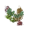
- EMDB-59866: Phosphorylated STING bound to adaptor protein complex 1 (processe... -
+ Open data

- Basic information
| Entry |  | |||||||||
|---|---|---|---|---|---|---|---|---|---|---|
| Title | Phosphorylated STING bound to adaptor protein complex 1 (processed for improved pSTING density) | |||||||||
 Map data Map data | ||||||||||
 Sample Sample |
| |||||||||
 Keywords Keywords | STING / innate immunity / TGN / AP-1 / IMMUNE SYSTEM | |||||||||
| Biological species |  Homo sapiens (human) / Homo sapiens (human) /   Mus musculus (house mouse) Mus musculus (house mouse) | |||||||||
| Method | single particle reconstruction / cryo EM / Resolution: 2.48 Å | |||||||||
 Authors Authors | Xu P / Ablasser A | |||||||||
| Funding support | European Union, 1 items
| |||||||||
 Citation Citation | Journal: Nature / Year: 2022 Title: Clathrin-associated AP-1 controls termination of STING signalling. Authors: Ying Liu / Pengbiao Xu / Sophie Rivara / Chong Liu / Jonathan Ricci / Xuefeng Ren / James H Hurley / Andrea Ablasser /   Abstract: Stimulator of interferon genes (STING) functions downstream of cyclic GMP-AMP synthase in DNA sensing or as a direct receptor for bacterial cyclic dinucleotides and small molecules to activate ...Stimulator of interferon genes (STING) functions downstream of cyclic GMP-AMP synthase in DNA sensing or as a direct receptor for bacterial cyclic dinucleotides and small molecules to activate immunity during infection, cancer and immunotherapy. Precise regulation of STING is essential to ensure balanced immune responses and prevent detrimental autoinflammation. After activation, STING, a transmembrane protein, traffics from the endoplasmic reticulum to the Golgi, where its phosphorylation by the protein kinase TBK1 enables signal transduction. The mechanism that ends STING signalling at the Golgi remains unknown. Here we show that adaptor protein complex 1 (AP-1) controls the termination of STING-dependent immune activation. We find that AP-1 sorts phosphorylated STING into clathrin-coated transport vesicles for delivery to the endolysosomal system, where STING is degraded. We identify a highly conserved dileucine motif in the cytosolic C-terminal tail (CTT) of STING that, together with TBK1-dependent CTT phosphorylation, dictates the AP-1 engagement of STING. A cryo-electron microscopy structure of AP-1 in complex with phosphorylated STING explains the enhanced recognition of TBK1-activated STING. We show that suppression of AP-1 exacerbates STING-induced immune responses. Our results reveal a structural mechanism of negative regulation of STING and establish that the initiation of signalling is inextricably associated with its termination to enable transient activation of immunity. | |||||||||
| History |
|
- Structure visualization
| Supplemental images |
|---|

- Downloads & links
-EMDB archive
| Map data | emd_59866.map.gz | 49.8 MB |  EMDB map data format EMDB map data format | |
|---|---|---|---|---|
| Header (meta data) | emd-59866-v30.xmlemd-59866.xml | 20.9 KB 20.9 KB | Display Display | EMDB header |
| FSC (resolution estimation) | emd_59866_fsc.xml | 9.9 KB | Display | FSC data file |
| Images |  emd_59866.png emd_59866.png | 106.9 KB | ||
| Filedesc metadata | emd-59866.cif.gz | 6.5 KB | ||
| Others | emd_59866_half_map_1.map.gzemd_59866_half_map_2.map.gz | 88 MB 88 MB | ||
| Archive directory |  https://data.pdbj.org/pub/emdb/structures/EMD-59866ftp://data.pdbj.org/pub/emdb/structures/EMD-59866 https://data.pdbj.org/pub/emdb/structures/EMD-59866ftp://data.pdbj.org/pub/emdb/structures/EMD-59866 | HTTPS FTP |
-Related structure data

-Links
| EMDB pages | EMDB (EBI/PDBe) / EMDataResource |
|---|
-Map
| File | Download / File: emd_59866.map.gz / Format: CCP4 / Size: 103 MB / Type: IMAGE STORED AS FLOATING POINT NUMBER (4 BYTES) | ||||||||||||||||||||||||||||||||||||
|---|---|---|---|---|---|---|---|---|---|---|---|---|---|---|---|---|---|---|---|---|---|---|---|---|---|---|---|---|---|---|---|---|---|---|---|---|---|
| Projections & slices | Image control
Images are generated by Spider. | ||||||||||||||||||||||||||||||||||||
| Voxel size | X=Y=Z: 0.9 Å | ||||||||||||||||||||||||||||||||||||
| Density |
| ||||||||||||||||||||||||||||||||||||
| Symmetry | Space group: 1 | ||||||||||||||||||||||||||||||||||||
| Details | EMDB XML:
|
 Z (Sec.)
 Y (Row.)
 X (Col.)


-Supplemental data
-Half map: #1
| File | emd_59866_half_map_1.map | ||||||||||||
|---|---|---|---|---|---|---|---|---|---|---|---|---|---|
| Projections & Slices |
| ||||||||||||
| Density Histograms |


-Half map: #2
| File | emd_59866_half_map_2.map | ||||||||||||
|---|---|---|---|---|---|---|---|---|---|---|---|---|---|
| Projections & Slices |
| ||||||||||||
| Density Histograms |


- Sample components
-Entire : phospho-STING binding to adaptor protein complex-1
| Entire | Name: phospho-STING binding to adaptor protein complex-1 |
|---|---|
| Components |
|
-Supramolecule #1: phospho-STING binding to adaptor protein complex-1
| Supramolecule | Name: phospho-STING binding to adaptor protein complex-1 / type: complex / ID: 1 / Parent: 0 / Macromolecule list: all |
|---|---|
| Source (natural) | Organism: Homo sapiens (human) |
-Macromolecule #1: AP-1 complex subunit beta-1
| Macromolecule | Name: AP-1 complex subunit beta-1 / type: protein_or_peptide / ID: 1 / Number of copies: 1 / Enantiomer: LEVO |
|---|---|
| Source (natural) | Organism: Homo sapiens (human) |
| Molecular weight | Theoretical: 66.008422 KDa |
| Recombinant expression | Organism:   Escherichia coli BL21(DE3) (bacteria) Escherichia coli BL21(DE3) (bacteria) |
| Sequence | String: MTDSKYFTTT KKGEIFELKA ELNSDKKEKK KEAVKKVIAS MTVGKDVSAL FPDVVNCMQT DNLELKKLVY LYLMNYAKSQ PDMAIMAVN TFVKDCEDPN PLIRALAVRT MGCIRVDKIT EYLCEPLRKC LKDEDPYVRK TAAVCVAKLH DINAQLVEDQ G FLDTLKDL ...String: MTDSKYFTTT KKGEIFELKA ELNSDKKEKK KEAVKKVIAS MTVGKDVSAL FPDVVNCMQT DNLELKKLVY LYLMNYAKSQ PDMAIMAVN TFVKDCEDPN PLIRALAVRT MGCIRVDKIT EYLCEPLRKC LKDEDPYVRK TAAVCVAKLH DINAQLVEDQ G FLDTLKDL ISDSNPMVVA NAVAALSEIA ESHPSSNLLD LNPQSINKLL TALNECTEWG QIFILDCLAN YMPKDDREAQ SI CERVTPR LSHANSAVVL SAVKVLMKFM EMLSKDLDYY GTLLKKLAPP LVTLLSAEPE LQYVALRNIN LIVQKRPEIL KHE MKVFFV KYNDPIYVKL EKLDIMIRLA SQANIAQVLA ELREYATEVD VDFVRKAVRA IGRCAIKVEQ SAERCVSTLL DLIQ TKVNY VVQEAIVVIK DIFRKYPNKY ESVIATLCEN LDSLDEPEAR AAMIWIVGEY AERIDNADEL LESFLEGFHD KSTQV QLQL LTAIVKLFLK KPTETQELVQ QVLSLATQDS DNPDLRDRGY IYWRLLSTDP VAAKEVVLAE KPLISEETDL IEPTLL DEL ICYIGTLASV YHKPPSAFVE G |
-Macromolecule #2: ADP-ribosylation factor 1
| Macromolecule | Name: ADP-ribosylation factor 1 / type: protein_or_peptide / ID: 2 / Number of copies: 2 / Enantiomer: LEVO |
|---|---|
| Source (natural) | Organism: Homo sapiens (human) |
| Molecular weight | Theoretical: 18.9366 KDa |
| Recombinant expression | Organism: Escherichia coli BL21(DE3) (bacteria) |
| Sequence | String: EMRILMVGLD AAGKTTILYK LKLGEIVTTI PTIGFNVETV EYKNISFTVW DVGGLDKIRP LWRHYFQNTQ GLIFVVDSND RERVNEARE ELMRMLAEDE LRDAVLLVFA NKQDLPNAMN AAEITDKLGL HSLRHRNWYI QATCATSGDG LYEGLDWLSN Q LRNQK |
-Macromolecule #3: AP-1 complex subunit gamma-1
| Macromolecule | Name: AP-1 complex subunit gamma-1 / type: protein_or_peptide / ID: 3 / Number of copies: 1 / Enantiomer: LEVO |
|---|---|
| Source (natural) | Organism: Mus musculus (house mouse) |
| Molecular weight | Theoretical: 67.399242 KDa |
| Recombinant expression | Organism: Escherichia coli BL21(DE3) (bacteria) |
| Sequence | String: MPAPIRLREL IRTIRTARTQ AEEREMIQKE CAAIRSSFRE EDNTYRCRNV AKLLYMHMLG YPAHFGQLEC LKLIASQKFT DKRIGYLGA MLLLDERQDV HLLMTNCIKN DLNHSTQFVQ GLALCTLGCM GSSEMCRDLA GEVEKLLKTS NSYLRKKAAL C AVHVIRKV ...String: MPAPIRLREL IRTIRTARTQ AEEREMIQKE CAAIRSSFRE EDNTYRCRNV AKLLYMHMLG YPAHFGQLEC LKLIASQKFT DKRIGYLGA MLLLDERQDV HLLMTNCIKN DLNHSTQFVQ GLALCTLGCM GSSEMCRDLA GEVEKLLKTS NSYLRKKAAL C AVHVIRKV PELMEMFLPA TKNLLNEKNH GVLHTSVVLL TEMCERSPDM LAHFRKLVPQ LVRILKNLIM SGYSPEHDVS GI SDPFLQV RILRLLRILG RNDDDSSEAM NDILAQVATN TETSKNVGNA ILYETVLTIM DIKSESGLRV LAINILGRFL LNN DKNIRY VALTSLLKTV QTDHNAVQRH RSTIVDCLKD LDVSIKRRAM ELSFALVNGN NIRGMMKELL YFLDSCEPEF KADC ASGIF LAAEKYAPSK RWHIDTIMRV LTTAGSYVRD DAVPNLIQLI TNSVEMHAYT VQRLYKAILG DYSQQPLVQV AAWCI GEYG DLLVSGQCEE EEPIQVTEDE VLDILESVLI SNMSTSVTRG YALTAIMKLS TRFTCTVNRI KKVVSIYGSS IDVELQ QRA VEYNALFKKY DHMRSALLER MPVMEKVTTN GP |
-Macromolecule #4: Stimulator of interferon genes protein
| Macromolecule | Name: Stimulator of interferon genes protein / type: protein_or_peptide / ID: 4 / Details: phospho-STING tail / Number of copies: 1 / Enantiomer: LEVO |
|---|---|
| Source (natural) | Organism: Homo sapiens (human) |
| Molecular weight | Theoretical: 1.065068 KDa |
| Recombinant expression | Organism: Escherichia coli BL21(DE3) (bacteria) |
| Sequence | String: QEPELLI(SEP)G |
-Macromolecule #5: AP-1 complex subunit mu-1
| Macromolecule | Name: AP-1 complex subunit mu-1 / type: protein_or_peptide / ID: 5 / Number of copies: 1 / Enantiomer: LEVO |
|---|---|
| Source (natural) | Organism: Mus musculus (house mouse) |
| Molecular weight | Theoretical: 48.60673 KDa |
| Recombinant expression | Organism: Escherichia coli BL21(DE3) (bacteria) |
| Sequence | String: MSASAVYVLD LKGKVLICRN YRGDVDMSEV EHFMPILMEK EEEGMLSPIL AHGGVRFMWI KHNNLYLVAT SKKNACVSLV FSFLYKVVQ VFSEYFKELE EESIRDNFVI IYELLDELMD FGYPQTTDSK ILQEYITQEG HKLETGAPRP PATVTNAVSW R SEGIKYRK ...String: MSASAVYVLD LKGKVLICRN YRGDVDMSEV EHFMPILMEK EEEGMLSPIL AHGGVRFMWI KHNNLYLVAT SKKNACVSLV FSFLYKVVQ VFSEYFKELE EESIRDNFVI IYELLDELMD FGYPQTTDSK ILQEYITQEG HKLETGAPRP PATVTNAVSW R SEGIKYRK NEVFLDVIEA VNLLVSANGN VLRSEIVGSI KMRVFLSGMP ELRLGLNDKV LFDNTGRGKS KSVELEDVKF HQ CVRLSRF ENDRTISFIP PDGEFELMSY RLNTHVKPLI WIESVIEKHS HSRIEYMVKA KSQFKRRSTA NNVEIHIPVP NDA DSPKFK TTVGSVKWVP ENSEIVWSVK SFPGGKEYLM RAHFGLPSVE AEDKEGKPPI SVKFEIPYFT TSGIQVRYLK IIEK SGYQA LPWVRYITQN GDYQLRTQ |
-Macromolecule #6: AP-1 complex subunit sigma-3
| Macromolecule | Name: AP-1 complex subunit sigma-3 / type: protein_or_peptide / ID: 6 / Number of copies: 1 / Enantiomer: LEVO |
|---|---|
| Source (natural) | Organism: Homo sapiens (human) |
| Molecular weight | Theoretical: 18.321338 KDa |
| Recombinant expression | Organism: Escherichia coli BL21(DE3) (bacteria) |
| Sequence | String: MIHFILLFSR QGKLRLQKWY ITLPDKERKK ITREIVQIIL SRGHRTSSFV DWKELKLVYK RYASLYFCCA IENQDNELLT LEIVHRYVE LLDKYFGNVC ELDIIFNFEK AYFILDEFII GGEIQETSKK IAVKAIEDSD MLQEVSTVCQ TMGER |
-Experimental details
-Structure determination
| Method | cryo EM |
|---|---|
 Processing Processing | single particle reconstruction |
| Aggregation state | particle |
-Sample preparation
| Concentration | 0.8 mg/mL |
|---|---|
| Buffer | pH: 7.4 / Details: PBS buffer |
| Grid | Model: Quantifoil R1.2/1.3 / Material: GOLD / Mesh: 300 / Support film - Material: CARBON / Support film - topology: HOLEY |
| Vitrification | Cryogen name: ETHANE |
- Electron microscopy
| Microscope | TFS KRIOS |
|---|---|
| Image recording | Film or detector model: FEI FALCON IV (4k x 4k) / Average electron dose: 60.0 e/Å2 |
| Electron beam | Acceleration voltage: 300 kV / Electron source:  FIELD EMISSION GUN FIELD EMISSION GUN |
| Electron optics | Illumination mode: FLOOD BEAM / Imaging mode: BRIGHT FIELD / Nominal defocus max: 1.8 µm / Nominal defocus min: 0.8 µm |
| Experimental equipment |  Model: Titan Krios / Image courtesy: FEI Company |

