ムービー
ムービー コントローラー


+ データを開く

- 基本情報
基本情報
| 登録情報 | データベース: EMDB / ID: EMD-5985 | |||||||||
|---|---|---|---|---|---|---|---|---|---|---|
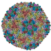
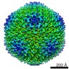
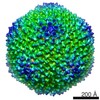
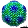
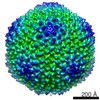
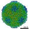
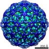
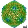
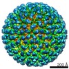
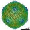
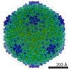
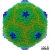
| タイトル | CryoEM of Bacteriophage PRD1 procapsid | |||||||||
 マップデータ マップデータ | Reconstruction of PRD1 procapsid without icosahedral symmetry imposition | |||||||||
 試料 試料 |
| |||||||||
 キーワード キーワード | Virus / Genome packaging | |||||||||
| 生物種 | Bacteriophage PRD1 procapsid | |||||||||
| 手法 | 単粒子再構成法 / クライオ電子顕微鏡法 / 解像度: 14.0 Å | |||||||||
 データ登録者 データ登録者 | Hong C / Oksanen HM / Liu X / Jakana J / Bamford DH / Chiu W | |||||||||
 引用 引用 | ジャーナル: PLoS Biol / 年: 2014 タイトル: A structural model of the genome packaging process in a membrane-containing double stranded DNA virus. 著者: Chuan Hong / Hanna M Oksanen / Xiangan Liu / Joanita Jakana / Dennis H Bamford / Wah Chiu /   要旨: Two crucial steps in the virus life cycle are genome encapsidation to form an infective virion and genome exit to infect the next host cell. In most icosahedral double-stranded (ds) DNA viruses, the ...Two crucial steps in the virus life cycle are genome encapsidation to form an infective virion and genome exit to infect the next host cell. In most icosahedral double-stranded (ds) DNA viruses, the viral genome enters and exits the capsid through a unique vertex. Internal membrane-containing viruses possess additional complexity as the genome must be translocated through the viral membrane bilayer. Here, we report the structure of the genome packaging complex with a membrane conduit essential for viral genome encapsidation in the tailless icosahedral membrane-containing bacteriophage PRD1. We utilize single particle electron cryo-microscopy (cryo-EM) and symmetry-free image reconstruction to determine structures of PRD1 virion, procapsid, and packaging deficient mutant particles. At the unique vertex of PRD1, the packaging complex replaces the regular 5-fold structure and crosses the lipid bilayer. These structures reveal that the packaging ATPase P9 and the packaging efficiency factor P6 form a dodecameric portal complex external to the membrane moiety, surrounded by ten major capsid protein P3 trimers. The viral transmembrane density at the special vertex is assigned to be a hexamer of heterodimer of proteins P20 and P22. The hexamer functions as a membrane conduit for the DNA and as a nucleating site for the unique vertex assembly. Our structures show a conformational alteration in the lipid membrane after the P9 and P6 are recruited to the virion. The P8-genome complex is then packaged into the procapsid through the unique vertex while the genome terminal protein P8 functions as a valve that closes the channel once the genome is inside. Comparing mature virion, procapsid, and mutant particle structures led us to propose an assembly pathway for the genome packaging apparatus in the PRD1 virion. | |||||||||
| 履歴 |
|
- 構造の表示
構造の表示
| ムービー |
 ムービービューア ムービービューア |
|---|---|
| 構造ビューア | EMマップ: SurfViewMolmilJmol/JSmol |
| 添付画像 |

- ダウンロードとリンク
-EMDBアーカイブ
| マップデータ | emd_5985.map.gz | 158.1 MB | EMDBマップデータ形式 | |
|---|---|---|---|---|
| ヘッダ (付随情報) | emd-5985-v30.xmlemd-5985.xml | 11.3 KB 11.3 KB | 表示 表示 | EMDBヘッダ |
| 画像 |  400_5985.gif 400_5985.gif 80_5985.gif 80_5985.gif | 113.6 KB 6.7 KB | ||
| アーカイブディレクトリ |  http://ftp.pdbj.org/pub/emdb/structures/EMD-5985ftp://ftp.pdbj.org/pub/emdb/structures/EMD-5985 http://ftp.pdbj.org/pub/emdb/structures/EMD-5985ftp://ftp.pdbj.org/pub/emdb/structures/EMD-5985 | HTTPS FTP |
-関連構造データ

-リンク
| EMDBのページ | EMDB (EBI/PDBe) / EMDataResource |
|---|
-マップ
| ファイル | ダウンロード / ファイル: emd_5985.map.gz / 形式: CCP4 / 大きさ: 804.7 MB / タイプ: IMAGE STORED AS FLOATING POINT NUMBER (4 BYTES) | ||||||||||||||||||||||||||||||||||||||||||||||||||||||||||||||||||||
|---|---|---|---|---|---|---|---|---|---|---|---|---|---|---|---|---|---|---|---|---|---|---|---|---|---|---|---|---|---|---|---|---|---|---|---|---|---|---|---|---|---|---|---|---|---|---|---|---|---|---|---|---|---|---|---|---|---|---|---|---|---|---|---|---|---|---|---|---|---|
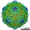
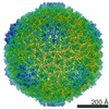
| 注釈 | Reconstruction of PRD1 procapsid without icosahedral symmetry imposition | ||||||||||||||||||||||||||||||||||||||||||||||||||||||||||||||||||||
| 投影像・断面図 | 画像のコントロール
画像は Spider により作成 | ||||||||||||||||||||||||||||||||||||||||||||||||||||||||||||||||||||
| ボクセルのサイズ | X=Y=Z: 1.42 Å | ||||||||||||||||||||||||||||||||||||||||||||||||||||||||||||||||||||
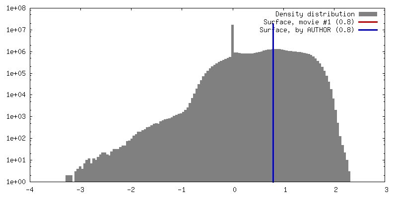
| 密度 |
| ||||||||||||||||||||||||||||||||||||||||||||||||||||||||||||||||||||
| 対称性 | 空間群: 1 | ||||||||||||||||||||||||||||||||||||||||||||||||||||||||||||||||||||
| 詳細 | EMDB XML:
CCP4マップ ヘッダ情報:
| ||||||||||||||||||||||||||||||||||||||||||||||||||||||||||||||||||||
 Z (Sec.)
 Y (Row.)
 X (Col.)


-添付データ
- 試料の構成要素
-全体 : Bacteriophage PRD1 procapsid
| 全体 | 名称: Bacteriophage PRD1 procapsid |
|---|---|
| 要素 |
|
-超分子 #1000: Bacteriophage PRD1 procapsid
| 超分子 | 名称: Bacteriophage PRD1 procapsid / タイプ: sample / ID: 1000 / 詳細: The genome DNA was not packaged. / 集合状態: Icosahedral Virus / Number unique components: 1 |
|---|---|
| 分子量 | 実験値: 66 MDa / 理論値: 66 MDa / 手法: Sedimentation |
-超分子 #1: Bacteriophage PRD1 procapsid
| 超分子 | 名称: Bacteriophage PRD1 procapsid / タイプ: virus / ID: 1 / 詳細: Packaging deficient procapsid / 生物種: Bacteriophage PRD1 procapsid / ウイルスタイプ: VIRUS-LIKE PARTICLE / ウイルス・単離状態: SPECIES / ウイルス・エンベロープ: No / ウイルス・中空状態: Yes |
|---|---|
| 宿主 | 生物種:  Salmonella enterica subsp. enterica serovar Typhimurium str. LT2 (サルモネラ菌) Salmonella enterica subsp. enterica serovar Typhimurium str. LT2 (サルモネラ菌)別称: BACTERIA(EUBACTERIA) |
| Host system | 生物種: Salmonella enterica subsp. enterica serovar Typhimurium str. LT2 (サルモネラ菌) |
| 分子量 | 実験値: 66 MDa / 理論値: 66 MDa |
| ウイルス殻 | Shell ID: 1 / 直径: 650 Å / T番号(三角分割数): 25 |
-実験情報
-構造解析
| 手法 | クライオ電子顕微鏡法 |
|---|---|
 解析 解析 | 単粒子再構成法 |
| 試料の集合状態 | particle |
-試料調製
| 濃度 | 10 mg/mL |
|---|---|
| 緩衝液 | pH: 7.2 / 詳細: 20 mM potassium phosphate, 1 mM MgCl2 |
| グリッド | 詳細: 400 mesh 1.2/1.3, plasma-cleaned |
| 凍結 | 凍結剤: ETHANE / チャンバー内湿度: 90 % / チャンバー内温度: 100 K / 装置: FEI VITROBOT MARK III / 手法: Blot for 2 seconds before plunging. |
- 電子顕微鏡法
| 顕微鏡 | JEOL 3200FSC |
|---|---|
| 温度 | 最低: 80 K / 最高: 90 K / 平均: 85 K |
| アライメント法 | Legacy - 非点収差: Objective lens astigmatism was corrected at 200,000 times magnification. |
| 特殊光学系 | エネルギーフィルター - 名称: JEOL Omega エネルギーフィルター - エネルギー下限: 0.0 eV エネルギーフィルター - エネルギー上限: 20.0 eV |
| 日付 | 2009年8月21日 |
| 撮影 | カテゴリ: CCD フィルム・検出器のモデル: GATAN ULTRASCAN 4000 (4k x 4k) デジタル化 - サンプリング間隔: 15 µm / 実像数: 340 / 平均電子線量: 20 e/Å2 / カメラ長: 150 |
| 電子線 | 加速電圧: 300 kV / 電子線源:  FIELD EMISSION GUN FIELD EMISSION GUN |
| 電子光学系 | 倍率(補正後): 106000 / 照射モード: FLOOD BEAM / 撮影モード: BRIGHT FIELD / Cs: 4.1 mm / 最大 デフォーカス(公称値): 3.5 µm / 最小 デフォーカス(公称値): 0.5 µm / 倍率(公称値): 80000 |
| 試料ステージ | 試料ホルダーモデル: JEOL 3200FSC CRYOHOLDER |
-画像解析
| 詳細 | No icosahedral symmetry was imposed. |
|---|---|
| CTF補正 | 詳細: Each frame |
| 最終 再構成 | アルゴリズム: OTHER / 解像度のタイプ: BY AUTHOR / 解像度: 14.0 Å / 解像度の算出法: OTHER / ソフトウェア - 名称: MPSA, EMAN, EMAN2 / 使用した粒子像数: 4300 |
-原子モデル構築 1
| 初期モデル | PDB ID: |
|---|---|
| ソフトウェア | 名称: Chimera |
| 精密化 | 空間: REAL / プロトコル: RIGID BODY FIT |