 Movie
Movie Controller

[English] 日本語
 Yorodumi
- EMDB-5805: Cryo-EM map of the Drosophila 80S Ribosome (control map for 80S-F... -
+ Open data

- Basic information
| Entry | Database: EMDB / ID: EMD-5805 | |||||||||
|---|---|---|---|---|---|---|---|---|---|---|
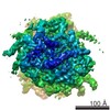
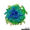
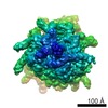
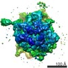
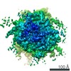
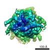
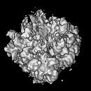
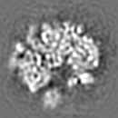
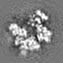
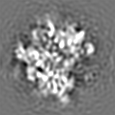
| Title | Cryo-EM map of the Drosophila 80S Ribosome (control map for 80S-FMRP Complex map) | |||||||||
 Map data Map data | Cryo-EM reconstruction of Drosophila 80S ribosome | |||||||||
 Sample Sample |
| |||||||||
 Keywords Keywords | Drosophila 80S FMRP | |||||||||
| Biological species |  | |||||||||
| Method | single particle reconstruction / cryo EM / Resolution: 11.2 Å | |||||||||
 Authors Authors | Chen E / Sharma M R / Shi X / Agrawal R K / Joseph S | |||||||||
 Citation Citation | Journal: Mol Cell / Year: 2014 Title: Fragile X mental retardation protein regulates translation by binding directly to the ribosome. Authors: Eileen Chen / Manjuli R Sharma / Xinying Shi / Rajendra K Agrawal / Simpson Joseph /  Abstract: Fragile X syndrome (FXS) is the most common form of inherited mental retardation, and it is caused by loss of function of the fragile X mental retardation protein (FMRP). FMRP is an RNA-binding ...Fragile X syndrome (FXS) is the most common form of inherited mental retardation, and it is caused by loss of function of the fragile X mental retardation protein (FMRP). FMRP is an RNA-binding protein that is involved in the translational regulation of several neuronal mRNAs. However, the precise mechanism of translational inhibition by FMRP is unknown. Here, we show that FMRP inhibits translation by binding directly to the L5 protein on the 80S ribosome. Furthermore, cryoelectron microscopic reconstruction of the 80S ribosome⋅FMRP complex shows that FMRP binds within the intersubunit space of the ribosome such that it would preclude the binding of tRNA and translation elongation factors on the ribosome. These findings suggest that FMRP inhibits translation by blocking the essential components of the translational machinery from binding to the ribosome. | |||||||||
| History |
|
- Structure visualization
| Movie |
 Movie viewer Movie viewer |
|---|---|
| Structure viewer | EM map: SurfViewMolmilJmol/JSmol |
| Supplemental images |

- Downloads & links
-EMDB archive
| Map data | emd_5805.map.gz | 7.8 MB | EMDB map data format | |
|---|---|---|---|---|
| Header (meta data) | emd-5805-v30.xmlemd-5805.xml | 10 KB 10 KB | Display Display | EMDB header |
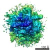
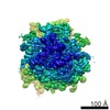
| Images | 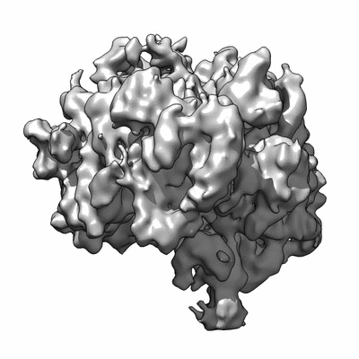 400_5805.gif 400_5805.gif 80_5805.gif 80_5805.gif | 37 KB 3.8 KB | ||
| Archive directory |  http://ftp.pdbj.org/pub/emdb/structures/EMD-5805ftp://ftp.pdbj.org/pub/emdb/structures/EMD-5805 http://ftp.pdbj.org/pub/emdb/structures/EMD-5805ftp://ftp.pdbj.org/pub/emdb/structures/EMD-5805 | HTTPS FTP |
-Validation report
| Summary document | emd_5805_validation.pdf.gz | 77.7 KB | Display | EMDB validaton report |
|---|---|---|---|---|
| Full document | emd_5805_full_validation.pdf.gz | 76.8 KB | Display | |
| Data in XML | emd_5805_validation.xml.gz | 493 B | Display | |
| Arichive directory | https://ftp.pdbj.org/pub/emdb/validation_reports/EMD-5805ftp://ftp.pdbj.org/pub/emdb/validation_reports/EMD-5805 | HTTPS FTP |
-Related structure data

-Links
| EMDB pages | EMDB (EBI/PDBe) / EMDataResource |
|---|---|
| Related items in Molecule of the Month |

-Map
| File | Download / File: emd_5805.map.gz / Format: CCP4 / Size: 8.2 MB / Type: IMAGE STORED AS FLOATING POINT NUMBER (4 BYTES) | ||||||||||||||||||||||||||||||||||||||||||||||||||||||||||||||||||||
|---|---|---|---|---|---|---|---|---|---|---|---|---|---|---|---|---|---|---|---|---|---|---|---|---|---|---|---|---|---|---|---|---|---|---|---|---|---|---|---|---|---|---|---|---|---|---|---|---|---|---|---|---|---|---|---|---|---|---|---|---|---|---|---|---|---|---|---|---|---|
| Annotation | Cryo-EM reconstruction of Drosophila 80S ribosome | ||||||||||||||||||||||||||||||||||||||||||||||||||||||||||||||||||||
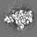
| Projections & slices | Image control
Images are generated by Spider. | ||||||||||||||||||||||||||||||||||||||||||||||||||||||||||||||||||||
| Voxel size | X=Y=Z: 2.78 Å | ||||||||||||||||||||||||||||||||||||||||||||||||||||||||||||||||||||
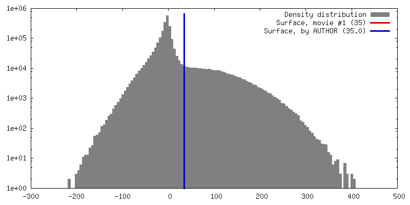
| Density |
| ||||||||||||||||||||||||||||||||||||||||||||||||||||||||||||||||||||
| Symmetry | Space group: 1 | ||||||||||||||||||||||||||||||||||||||||||||||||||||||||||||||||||||
| Details | EMDB XML:
CCP4 map header:
| ||||||||||||||||||||||||||||||||||||||||||||||||||||||||||||||||||||
 Z (Sec.)
 Y (Row.)
 X (Col.)


-Supplemental data
- Sample components
-Entire : Cryo-EM map of the Drosophila 80S Ribosome (control map for 80S-F...
| Entire | Name: Cryo-EM map of the Drosophila 80S Ribosome (control map for 80S-FMRP Complex map) |
|---|---|
| Components |
|
-Supramolecule #1000: Cryo-EM map of the Drosophila 80S Ribosome (control map for 80S-F...
| Supramolecule | Name: Cryo-EM map of the Drosophila 80S Ribosome (control map for 80S-FMRP Complex map) type: sample / ID: 1000 / Details: monodisperse / Number unique components: 1 |
|---|
-Supramolecule #1: 80S ribosome
| Supramolecule | Name: 80S ribosome / type: complex / ID: 1 / Recombinant expression: No / Database: NCBI / Ribosome-details: ribosome-eukaryote: ALL |
|---|---|
| Source (natural) | Organism: |
| Molecular weight | Experimental: 3 MDa / Theoretical: 3 MDa |
-Experimental details
-Structure determination
| Method | cryo EM |
|---|---|
 Processing Processing | single particle reconstruction |
| Aggregation state | particle |
-Sample preparation
| Concentration | 0.12 mg/mL |
|---|---|
| Buffer | pH: 7.7 Details: 50 mM KOAc, 50 mM Tris acetate, pH 7.7, 10 mM DTT, 5 mM Mg(OAc)2 |
| Grid | Details: 300 mesh Quantifoil with thin carbon support, glow discharged |
| Vitrification | Cryogen name: ETHANE / Chamber humidity: 85 % / Chamber temperature: 120 K / Instrument: FEI VITROBOT MARK II / Method: Blot for 6 seconds before plunging. |
- Electron microscopy
| Microscope | FEI TECNAI 20 |
|---|---|
| Temperature | Average: 120 K |
| Date | Dec 27, 2011 |
| Image recording | Category: FILM / Film or detector model: KODAK SO-163 FILM / Digitization - Scanner: ZEISS SCAI / Digitization - Sampling interval: 14 µm / Number real images: 102 / Average electron dose: 10 e/Å2 / Bits/pixel: 32 |
| Electron beam | Acceleration voltage: 200 kV / Electron source:  FIELD EMISSION GUN FIELD EMISSION GUN |
| Electron optics | Calibrated magnification: 50760 / Illumination mode: SPOT SCAN / Imaging mode: BRIGHT FIELD / Cs: 2 mm / Nominal defocus max: 3.5 µm / Nominal defocus min: 1.0 µm / Nominal magnification: 50000 |
| Sample stage | Specimen holder: liquid nitrogen-cooled / Specimen holder model: GATAN LIQUID NITROGEN |
