 ムービー
ムービー コントローラー


+ データを開く

- 基本情報
基本情報
| 登録情報 | データベース: EMDB / ID: EMD-5754 | |||||||||
|---|---|---|---|---|---|---|---|---|---|---|
| タイトル | Maximizing the potential of electron cryomicroscopy data collected using direct detectors | |||||||||
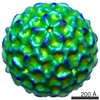
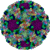
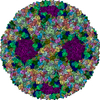
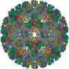
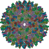
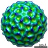
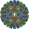
 マップデータ マップデータ | Reconstruction of mature STIV virion | |||||||||
 試料 試料 |
| |||||||||
 キーワード キーワード | Electron microscopy / Direct detectors / Near-atomic resolution / Sulfolobus turreted icosahedral virus | |||||||||
| 生物種 |    Sulfolobus turreted icosahedral virus (ウイルス) Sulfolobus turreted icosahedral virus (ウイルス) | |||||||||
| 手法 | 単粒子再構成法 / クライオ電子顕微鏡法 / 解像度: 5.8 Å | |||||||||
 データ登録者 データ登録者 | Veesler D / Campbell MG / Cheng A / Fu CY / Murez Z / Johnson JE / Potter CS / Carragher B | |||||||||
 引用 引用 | ジャーナル: J Struct Biol / 年: 2013 タイトル: Maximizing the potential of electron cryomicroscopy data collected using direct detectors. 著者: David Veesler / Melody G Campbell / Anchi Cheng / Chi-Yu Fu / Zachary Murez / John E Johnson / Clinton S Potter / Bridget Carragher /  要旨: Single-particle electron cryomicroscopy is undergoing a technical revolution due to the recent developments of direct detectors. These new recording devices detect electrons directly (i.e. without ...Single-particle electron cryomicroscopy is undergoing a technical revolution due to the recent developments of direct detectors. These new recording devices detect electrons directly (i.e. without conversion into light) and feature significantly improved detective quantum efficiencies and readout rates as compared to photographic films or CCDs. We evaluated here the potential of one such detector (Gatan K2 Summit) to enable the achievement of near-atomic resolution reconstructions of biological specimens when coupled to a widely used, mid-range transmission electron microscope (FEI TF20 Twin). Compensating for beam-induced motion and stage drift provided a 4.4Å resolution map of Sulfolobus turreted icosahedral virus (STIV), which we used as a test particle in this study. Several motion correction and dose fractionation procedures were explored and we describe their influence on the resolution of the final reconstruction. We also compared the quality of this data to that collected with a FEI Titan Krios microscope equipped with a Falcon I direct detector, which provides a benchmark for data collected using a high-end electron microscope. | |||||||||
| 履歴 |
|
- 構造の表示
構造の表示
| ムービー |
 ムービービューア ムービービューア |
|---|---|
| 構造ビューア | EMマップ: SurfViewMolmilJmol/JSmol |
| 添付画像 |
- ダウンロードとリンク
-EMDBアーカイブ
| マップデータ | emd_5754.map.gz | 3.3 GB | EMDBマップデータ形式 | |
|---|---|---|---|---|
| ヘッダ (付随情報) | emd-5754-v30.xmlemd-5754.xml | 10.2 KB 10.2 KB | 表示 表示 | EMDBヘッダ |
| 画像 |  400_5754.gif 400_5754.gif 80_5754.gif 80_5754.gif | 91.4 KB 5.5 KB | ||
| アーカイブディレクトリ |  http://ftp.pdbj.org/pub/emdb/structures/EMD-5754ftp://ftp.pdbj.org/pub/emdb/structures/EMD-5754 http://ftp.pdbj.org/pub/emdb/structures/EMD-5754ftp://ftp.pdbj.org/pub/emdb/structures/EMD-5754 | HTTPS FTP |
-検証レポート
| 文書・要旨 | emd_5754_validation.pdf.gz | 78.9 KB | 表示 | EMDB検証レポート |
|---|---|---|---|---|
| 文書・詳細版 | emd_5754_full_validation.pdf.gz | 78 KB | 表示 | |
| XML形式データ | emd_5754_validation.xml.gz | 494 B | 表示 | |
| アーカイブディレクトリ | https://ftp.pdbj.org/pub/emdb/validation_reports/EMD-5754ftp://ftp.pdbj.org/pub/emdb/validation_reports/EMD-5754 | HTTPS FTP |
-関連構造データ


-リンク
| EMDBのページ | EMDB (EBI/PDBe) / EMDataResource |
|---|
-マップ
| ファイル | ダウンロード / ファイル: emd_5754.map.gz / 形式: CCP4 / 大きさ: 3.9 GB / タイプ: IMAGE STORED AS FLOATING POINT NUMBER (4 BYTES) | ||||||||||||||||||||||||||||||||||||||||||||||||||||||||||||||||||||
|---|---|---|---|---|---|---|---|---|---|---|---|---|---|---|---|---|---|---|---|---|---|---|---|---|---|---|---|---|---|---|---|---|---|---|---|---|---|---|---|---|---|---|---|---|---|---|---|---|---|---|---|---|---|---|---|---|---|---|---|---|---|---|---|---|---|---|---|---|---|
| 注釈 | Reconstruction of mature STIV virion | ||||||||||||||||||||||||||||||||||||||||||||||||||||||||||||||||||||
| 投影像・断面図 | 画像のコントロール
画像は Spider により作成 | ||||||||||||||||||||||||||||||||||||||||||||||||||||||||||||||||||||
| ボクセルのサイズ | X=Y=Z: 1.21 Å | ||||||||||||||||||||||||||||||||||||||||||||||||||||||||||||||||||||
| 密度 |
| ||||||||||||||||||||||||||||||||||||||||||||||||||||||||||||||||||||
| 対称性 | 空間群: 1 | ||||||||||||||||||||||||||||||||||||||||||||||||||||||||||||||||||||
| 詳細 | EMDB XML:
CCP4マップ ヘッダ情報:
| ||||||||||||||||||||||||||||||||||||||||||||||||||||||||||||||||||||
 Z (Sec.)
 Y (Row.)
 X (Col.)


-添付データ
- 試料の構成要素
-全体 : Mature Sulfolobus Turreted Icosahedral Virus
| 全体 | 名称: Mature Sulfolobus Turreted Icosahedral Virus |
|---|---|
| 要素 |
|
-超分子 #1000: Mature Sulfolobus Turreted Icosahedral Virus
| 超分子 | 名称: Mature Sulfolobus Turreted Icosahedral Virus / タイプ: sample / ID: 1000 / 集合状態: icosahedral / Number unique components: 1 |
|---|---|
| 分子量 | 理論値: 75 MDa |
-超分子 #1: Sulfolobus turreted icosahedral virus
| 超分子 | 名称: Sulfolobus turreted icosahedral virus / タイプ: virus / ID: 1 / NCBI-ID: 269145 / 生物種: Sulfolobus turreted icosahedral virus / Sci species strain: YNPRC179 / データベース: NCBI / ウイルスタイプ: VIRION / ウイルス・単離状態: STRAIN / ウイルス・エンベロープ: Yes / ウイルス・中空状態: No |
|---|---|
| 宿主 | 生物種:  Sulfolobus solfataricus (古細菌) / 株: 2-2-12 / 別称: ARCHAEA Sulfolobus solfataricus (古細菌) / 株: 2-2-12 / 別称: ARCHAEA |
| 分子量 | 理論値: 75 MDa |
| ウイルス殻 | Shell ID: 1 / 直径: 730 Å |
-実験情報
-構造解析
| 手法 | クライオ電子顕微鏡法 |
|---|---|
 解析 解析 | 単粒子再構成法 |
| 試料の集合状態 | particle |
-試料調製
| 緩衝液 | pH: 3.5 詳細: 23 mM KH2PO4, 19 mM (NH4)2SO4, 1 mM MgSO4, 2 mM CaCl2 |
|---|---|
| グリッド | 詳細: plasma cleaned C-flat holey carbon grids (CF-1.2/1.3, Protochips) |
| 凍結 | 凍結剤: ETHANE / チャンバー内温度: 94 K / 装置: GATAN CRYOPLUNGE 3 / 詳細: Vitrification was carried out at room temperature. / 手法: Blot for 3 seconds before plunging. |
- 電子顕微鏡法
| 顕微鏡 | FEI TECNAI F20 |
|---|---|
| 温度 | 平均: 90 K |
| アライメント法 | Legacy - 非点収差: Objective lens astigmatism was corrected at 41322 times magnification |
| 日付 | 2012年11月30日 |
| 撮影 | カテゴリ: CCD / フィルム・検出器のモデル: GATAN K2 (4k x 4k) / 実像数: 754 / 平均電子線量: 22 e/Å2 詳細: Every movie is composed of sixteen frames recorded by the direct electron detector. |
| 電子線 | 加速電圧: 200 kV / 電子線源:  FIELD EMISSION GUN FIELD EMISSION GUN |
| 電子光学系 | 倍率(補正後): 41322 / 照射モード: FLOOD BEAM / 撮影モード: BRIGHT FIELD / Cs: 2.0 mm / 最大 デフォーカス(公称値): 3.7 µm / 最小 デフォーカス(公称値): 0.45 µm |
| 試料ステージ | 試料ホルダー: Nitrogen cooled / 試料ホルダーモデル: GATAN LIQUID NITROGEN |
| 実験機器 |  モデル: Tecnai F20 / 画像提供: FEI Company |
-画像解析
| 詳細 | The final reconstruction was sharpened with a negative temperature factor of 650 A^2. |
|---|---|
| CTF補正 | 詳細: Each particle |
| 最終 再構成 | アルゴリズム: OTHER / 解像度のタイプ: BY AUTHOR / 解像度: 5.8 Å / 解像度の算出法: OTHER / ソフトウェア - 名称: Frealign 詳細: The final reconstruction was computed using the first ten frames of each movie and sharpened with a negative temperature factor of 650 A^2. 使用した粒子像数: 4446 |
