 ムービー
ムービー コントローラー


+ データを開く

- 基本情報
基本情報
| 登録情報 | データベース: EMDB / ID: EMD-5718 | |||||||||
|---|---|---|---|---|---|---|---|---|---|---|
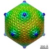
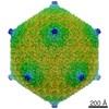
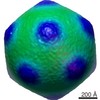
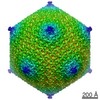
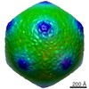
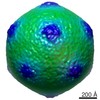
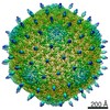
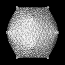
| タイトル | The structure of Sinorhizobium meliloti phage phiM12, a novel T=19 icosahedral phage that is the founder of a new group of T4-like phages | |||||||||
 マップデータ マップデータ | Cryo-EM reconstruction of phiM12, emptied of DNA | |||||||||
 試料 試料 |
| |||||||||
 キーワード キーワード | bacteriophage / phiM12 / Sinorhizobium meliloti / T=19 / rhizobia | |||||||||
| 生物種 |  Sinorhizobium phage phiM12 (ファージ) Sinorhizobium phage phiM12 (ファージ) | |||||||||
| 手法 | 単粒子再構成法 / クライオ電子顕微鏡法 / 解像度: 13.0 Å | |||||||||
 データ登録者 データ登録者 | Stroupe ME / Brewer TE / Sousa DR / Jones KM | |||||||||
 引用 引用 | ジャーナル: Virology / 年: 2014 タイトル: The structure of Sinorhizobium meliloti phage ΦM12, which has a novel T=19l triangulation number and is the founder of a new group of T4-superfamily phages. 著者: M Elizabeth Stroupe / Tess E Brewer / Duncan R Sousa / Kathryn M Jones /  要旨: ΦM12 is the first example of a T=19l geometry capsid, encapsulating the recently sequenced genome. Here, we present structures determined by cryo-EM of full and empty capsids. The structure reveals ...ΦM12 is the first example of a T=19l geometry capsid, encapsulating the recently sequenced genome. Here, we present structures determined by cryo-EM of full and empty capsids. The structure reveals the pattern for assembly of 1140 HK97-like capsid proteins, pointing to interactions at the pseudo 3-fold symmetry axes that hold together the asymmetric unit. The particular smooth surface of the capsid, along with a lack of accessory coat proteins encoded by the genome, suggest that this interface is the primary mechanism for capsid assembly. Two-dimensional averages of the tail, including the neck and baseplate, reveal that ΦM12 has a relatively narrow neck that attaches the tail to the capsid, as well as a three-layer baseplate. When free from DNA, the icosahedral edges expand by about 5nm, while the vertices stay at the same position, forming a similarly smooth, but bowed, T=19l icosahedral capsid. | |||||||||
| 履歴 |
|
- 構造の表示
構造の表示
| ムービー |
 ムービービューア ムービービューア |
|---|---|
| 構造ビューア | EMマップ: SurfViewMolmilJmol/JSmol |
| 添付画像 |
- ダウンロードとリンク
-EMDBアーカイブ
| マップデータ | emd_5718.map.gz | 19.1 MB | EMDBマップデータ形式 | |
|---|---|---|---|---|
| ヘッダ (付随情報) | emd-5718-v30.xmlemd-5718.xml | 9.3 KB 9.3 KB | 表示 表示 | EMDBヘッダ |
| 画像 |  400_5718.gif 400_5718.gif 80_5718.gif 80_5718.gif | 79.2 KB 5.2 KB | ||
| アーカイブディレクトリ |  http://ftp.pdbj.org/pub/emdb/structures/EMD-5718ftp://ftp.pdbj.org/pub/emdb/structures/EMD-5718 http://ftp.pdbj.org/pub/emdb/structures/EMD-5718ftp://ftp.pdbj.org/pub/emdb/structures/EMD-5718 | HTTPS FTP |
-検証レポート
| 文書・要旨 | emd_5718_validation.pdf.gz | 78.7 KB | 表示 | EMDB検証レポート |
|---|---|---|---|---|
| 文書・詳細版 | emd_5718_full_validation.pdf.gz | 77.8 KB | 表示 | |
| XML形式データ | emd_5718_validation.xml.gz | 494 B | 表示 | |
| アーカイブディレクトリ | https://ftp.pdbj.org/pub/emdb/validation_reports/EMD-5718ftp://ftp.pdbj.org/pub/emdb/validation_reports/EMD-5718 | HTTPS FTP |
-関連構造データ

-リンク
| EMDBのページ | EMDB (EBI/PDBe) / EMDataResource |
|---|
-マップ
| ファイル | ダウンロード / ファイル: emd_5718.map.gz / 形式: CCP4 / 大きさ: 41.9 MB / タイプ: IMAGE STORED AS FLOATING POINT NUMBER (4 BYTES) | ||||||||||||||||||||||||||||||||||||||||||||||||||||||||||||||||||||
|---|---|---|---|---|---|---|---|---|---|---|---|---|---|---|---|---|---|---|---|---|---|---|---|---|---|---|---|---|---|---|---|---|---|---|---|---|---|---|---|---|---|---|---|---|---|---|---|---|---|---|---|---|---|---|---|---|---|---|---|---|---|---|---|---|---|---|---|---|---|
| 注釈 | Cryo-EM reconstruction of phiM12, emptied of DNA | ||||||||||||||||||||||||||||||||||||||||||||||||||||||||||||||||||||
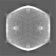
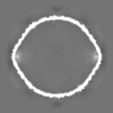
| 投影像・断面図 | 画像のコントロール
画像は Spider により作成 | ||||||||||||||||||||||||||||||||||||||||||||||||||||||||||||||||||||
| ボクセルのサイズ | X=Y=Z: 5.4 Å | ||||||||||||||||||||||||||||||||||||||||||||||||||||||||||||||||||||
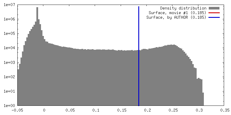
| 密度 |
| ||||||||||||||||||||||||||||||||||||||||||||||||||||||||||||||||||||
| 対称性 | 空間群: 1 | ||||||||||||||||||||||||||||||||||||||||||||||||||||||||||||||||||||
| 詳細 | EMDB XML:
CCP4マップ ヘッダ情報:
| ||||||||||||||||||||||||||||||||||||||||||||||||||||||||||||||||||||
 Z (Sec.)
 Y (Row.)
 X (Col.)


-添付データ
- 試料の構成要素
-全体 : Empty T=19 icosahedral shell from phiM12, a bacteriophage of Sino...
| 全体 | 名称: Empty T=19 icosahedral shell from phiM12, a bacteriophage of Sinorhizobium meliloti |
|---|---|
| 要素 |
|
-超分子 #1000: Empty T=19 icosahedral shell from phiM12, a bacteriophage of Sino...
| 超分子 | 名称: Empty T=19 icosahedral shell from phiM12, a bacteriophage of Sinorhizobium meliloti タイプ: sample / ID: 1000 / 集合状態: icosahedral / Number unique components: 1 |
|---|
-超分子 #1: Sinorhizobium phage phiM12
| 超分子 | 名称: Sinorhizobium phage phiM12 / タイプ: virus / ID: 1 / NCBI-ID: 1357423 / 生物種: Sinorhizobium phage phiM12 / Sci species strain: phiM12 / データベース: NCBI / ウイルスタイプ: VIRION / ウイルス・単離状態: STRAIN / ウイルス・エンベロープ: No / ウイルス・中空状態: No |
|---|---|
| 宿主 | 生物種:  Sinorhizobium meliloti (根粒菌) / 株: 1021 / 別称: BACTERIA(EUBACTERIA) Sinorhizobium meliloti (根粒菌) / 株: 1021 / 別称: BACTERIA(EUBACTERIA) |
| ウイルス殻 | Shell ID: 1 / 名称: 1 / 直径: 1000 Å / T番号(三角分割数): 19 |
-実験情報
-構造解析
| 手法 | クライオ電子顕微鏡法 |
|---|---|
 解析 解析 | 単粒子再構成法 |
| 試料の集合状態 | particle |
-試料調製
| 緩衝液 | pH: 7 / 詳細: 10 mM Na2HPO4, 1 mM MgSO4 |
|---|---|
| グリッド | 詳細: 200 mesh 2/2 Quantifoil |
| 凍結 | 凍結剤: ETHANE / チャンバー内湿度: 100 % / チャンバー内温度: 120 K / 装置: FEI VITROBOT MARK IV / 手法: blot 4 seconds |
- 電子顕微鏡法
| 顕微鏡 | FEI TITAN KRIOS |
|---|---|
| 日付 | 2013年5月13日 |
| 撮影 | カテゴリ: CCD フィルム・検出器のモデル: GATAN ULTRASCAN 4000 (4k x 4k) 実像数: 1000 / 平均電子線量: 15 e/Å2 |
| Tilt angle min | 0 |
| Tilt angle max | 0 |
| 電子線 | 加速電圧: 120 kV / 電子線源:  FIELD EMISSION GUN FIELD EMISSION GUN |
| 電子光学系 | 倍率(補正後): 65555 / 照射モード: FLOOD BEAM / 撮影モード: BRIGHT FIELD / 最大 デフォーカス(公称値): 3.5 µm / 最小 デフォーカス(公称値): 1.5 µm / 倍率(公称値): 59000 |
| 試料ステージ | 試料ホルダーモデル: FEI TITAN KRIOS AUTOGRID HOLDER |
| 実験機器 |  モデル: Titan Krios / 画像提供: FEI Company |
-画像解析
| 詳細 | Images were selected by hand; initial model determined in EMAN and refined in EMAN and Frealign |
|---|---|
| CTF補正 | 詳細: micrograph |
| 最終 再構成 | アルゴリズム: OTHER / 解像度のタイプ: BY AUTHOR / 解像度: 13.0 Å / 解像度の算出法: FSC 0.143 CUT-OFF / ソフトウェア - 名称: EMAN, FREALIGN / 使用した粒子像数: 2038 |
