 Movie
Movie Controller


+ Open data

- Basic information
| Entry | Database: EMDB / ID: EMD-7855 | ||||||||||||||||||
|---|---|---|---|---|---|---|---|---|---|---|---|---|---|---|---|---|---|---|---|
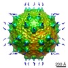
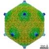
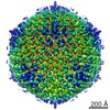
| Title | Carboxysome subtomogram averaging of size group 106 nm | ||||||||||||||||||
 Map data Map data | Subtomogram averaging of carboxysome size group 106 nm | ||||||||||||||||||
 Sample Sample |
| ||||||||||||||||||
| Biological species |  Synechococcus sp. WH 8109 (bacteria) Synechococcus sp. WH 8109 (bacteria) | ||||||||||||||||||
| Method | subtomogram averaging / cryo EM / Resolution: 40.0 Å | ||||||||||||||||||
 Authors Authors | Dai W / Chen M / Chiu W | ||||||||||||||||||
| Funding support |  United States, 5 items United States, 5 items
| ||||||||||||||||||
 Citation Citation | Journal: J Mol Biol / Year: 2018 Title: Visualizing Individual RuBisCO and Its Assembly into Carboxysomes in Marine Cyanobacteria by Cryo-Electron Tomography. Authors: Wei Dai / Muyuan Chen / Christopher Myers / Steven J Ludtke / B Montgomery Pettitt / Jonathan A King / Michael F Schmid / Wah Chiu / Abstract: Cyanobacteria are photosynthetic organisms responsible for ~25% of the organic carbon fixation on earth. A key step in carbon fixation is catalyzed by ribulose bisphosphate carboxylase/oxygenase ...Cyanobacteria are photosynthetic organisms responsible for ~25% of the organic carbon fixation on earth. A key step in carbon fixation is catalyzed by ribulose bisphosphate carboxylase/oxygenase (RuBisCO), the most abundant enzyme in the biosphere. Applying Zernike phase-contrast electron cryo-tomography and automated annotation, we identified individual RuBisCO molecules and their assembly intermediates leading to the formation of carboxysomes inside Syn5 cyanophage infected cyanobacteria Synechococcus sp. WH8109 cells. Surprisingly, more RuBisCO molecules were found to be present as cytosolic free-standing complexes or clusters than as packaged assemblies inside carboxysomes. Cytosolic RuBisCO clusters and partially assembled carboxysomes identified in the cell tomograms support a concurrent assembly model involving both the protein shell and the enclosed RuBisCO. In mature carboxysomes, RuBisCO is neither randomly nor strictly icosahedrally packed within protein shells of variable sizes. A time-averaged molecular dynamics simulation showed a semi-liquid probability distribution of the RuBisCO in carboxysomes and correlated well with carboxysome subtomogram averages. Our structural observations reveal the various stages of RuBisCO assemblies, which could be important for understanding cellular function. | ||||||||||||||||||
| History |
|
- Structure visualization
| Movie |
 Movie viewer Movie viewer |
|---|---|
| Structure viewer | EM map: SurfViewMolmilJmol/JSmol |
| Supplemental images |

- Downloads & links
-EMDB archive
| Map data | emd_7855.map.gz | 3.7 MB | EMDB map data format | |
|---|---|---|---|---|
| Header (meta data) | emd-7855-v30.xmlemd-7855.xml | 13.4 KB 13.4 KB | Display Display | EMDB header |
| Images | 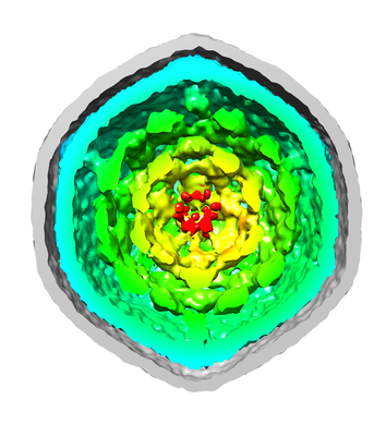 emd_7855.png emd_7855.png | 119.2 KB | ||
| Archive directory |  http://ftp.pdbj.org/pub/emdb/structures/EMD-7855ftp://ftp.pdbj.org/pub/emdb/structures/EMD-7855 http://ftp.pdbj.org/pub/emdb/structures/EMD-7855ftp://ftp.pdbj.org/pub/emdb/structures/EMD-7855 | HTTPS FTP |
-Related structure data


-Links
| EMDB pages | EMDB (EBI/PDBe) / EMDataResource |
|---|
-Map
| File | Download / File: emd_7855.map.gz / Format: CCP4 / Size: 38.4 MB / Type: IMAGE STORED AS FLOATING POINT NUMBER (4 BYTES) | ||||||||||||||||||||||||||||||||||||||||||||||||||||||||||||||||||||
|---|---|---|---|---|---|---|---|---|---|---|---|---|---|---|---|---|---|---|---|---|---|---|---|---|---|---|---|---|---|---|---|---|---|---|---|---|---|---|---|---|---|---|---|---|---|---|---|---|---|---|---|---|---|---|---|---|---|---|---|---|---|---|---|---|---|---|---|---|---|
| Annotation | Subtomogram averaging of carboxysome size group 106 nm | ||||||||||||||||||||||||||||||||||||||||||||||||||||||||||||||||||||
| Projections & slices | Image control
Images are generated by Spider. | ||||||||||||||||||||||||||||||||||||||||||||||||||||||||||||||||||||
| Voxel size | X=Y=Z: 9.04 Å | ||||||||||||||||||||||||||||||||||||||||||||||||||||||||||||||||||||
| Density |
| ||||||||||||||||||||||||||||||||||||||||||||||||||||||||||||||||||||
| Symmetry | Space group: 1 | ||||||||||||||||||||||||||||||||||||||||||||||||||||||||||||||||||||
| Details | EMDB XML:
CCP4 map header:
| ||||||||||||||||||||||||||||||||||||||||||||||||||||||||||||||||||||
 Z (Sec.)
 Y (Row.)
 X (Col.)


-Supplemental data
- Sample components
-Entire : carboxysome
| Entire | Name: carboxysome |
|---|---|
| Components |
|
-Supramolecule #1: carboxysome
| Supramolecule | Name: carboxysome / type: organelle_or_cellular_component / ID: 1 / Parent: 0 Details: carboxysome from cyanobacteria Synechococcus sp. WH8109 |
|---|---|
| Source (natural) | Organism: Synechococcus sp. WH 8109 (bacteria) / Organelle: carboxysome / Location in cell: cytoplasm |
-Experimental details
-Structure determination
| Method | cryo EM |
|---|---|
 Processing Processing | subtomogram averaging |
| Aggregation state | particle |
-Sample preparation
| Buffer | pH: 8 / Component - Name: Artificial sea water Details: Solutions were made fresh and autoclaved before use. |
|---|---|
| Grid | Model: Quantifoil R2/1 / Material: COPPER / Mesh: 200 / Support film - Material: CARBON / Support film - topology: HOLEY / Pretreatment - Type: GLOW DISCHARGE / Pretreatment - Atmosphere: AIR |
| Vitrification | Cryogen name: ETHANE / Chamber humidity: 90 % / Chamber temperature: 298 K / Instrument: FEI VITROBOT MARK IV / Details: Blot for 2 seconds before plunging. |
| Details | Carboxysome in Synechococcus sp. WH8109 cells |
- Electron microscopy
| Microscope | JEOL 2200FS |
|---|---|
| Temperature | Min: 70.0 K |
| Specialist optics | Phase plate: ZERNIKE PHASE PLATE / Energy filter - Name: In-column Omega Filter / Energy filter - Lower energy threshold: 0 eV / Energy filter - Upper energy threshold: 20 eV |
| Image recording | Film or detector model: GATAN ULTRASCAN 4000 (4k x 4k) / Average exposure time: 2.0 sec. / Average electron dose: 0.5 e/Å2 |
| Electron beam | Acceleration voltage: 200 kV / Electron source:  FIELD EMISSION GUN FIELD EMISSION GUN |
| Electron optics | C2 aperture diameter: 100.0 µm / Illumination mode: OTHER / Imaging mode: BRIGHT FIELD / Cs: 4.1 mm / Nominal defocus max: 0.0 µm / Nominal defocus min: 0.0 µm / Nominal magnification: 25000 |
| Sample stage | Specimen holder model: GATAN 626 SINGLE TILT LIQUID NITROGEN CRYO TRANSFER HOLDER Cooling holder cryogen: NITROGEN |
-Image processing
| Final reconstruction | Number classes used: 1 / Applied symmetry - Point group: I (icosahedral) / Algorithm: FOURIER SPACE / Resolution.type: BY AUTHOR / Resolution: 40.0 Å / Resolution method: FSC 0.5 CUT-OFF / Software - Name: EMAN2 (ver. 2.21) / Number subtomograms used: 28 |
|---|---|
| Extraction | Number tomograms: 56 / Number images used: 198 / Reference model: none / Method: carboxysome subvolumes selected manually / Software - Name: EMAN2 (ver. 2.21a) |
| Final 3D classification | Number classes: 2 / Details: Icosahedral/non-Icosahedral carboxysomes |
| Final angle assignment | Type: OTHER / Software - Name: EMAN2 (ver. 2.21) / Details: Subtomogram alignment and averaging in EMAN2 |
