 Movie
Movie Controller


+ Open data

- Basic information
| Entry | Database: EMDB / ID: EMD-1281 | |||||||||
|---|---|---|---|---|---|---|---|---|---|---|
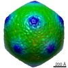
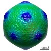
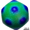
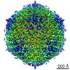
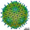
| Title | Determinants of bacteriophage phi29 head morphology. | |||||||||
 Map data Map data | This map is a cryo-EM 3D reconstruction of a fibered, T=4 isometric variant of bacteriophage phi29 | |||||||||
 Sample Sample |
| |||||||||
| Biological species |   Bacillus phage phi29 (virus) Bacillus phage phi29 (virus) | |||||||||
| Method | single particle reconstruction / cryo EM / Resolution: 20.0 Å | |||||||||
 Authors Authors | Choi KH / Morais MC / Anderson DL / Rossmann MG | |||||||||
 Citation Citation | Journal: Structure / Year: 2006 Title: Determinants of bacteriophage phi29 head morphology. Authors: Kyung H Choi / Marc C Morais / Dwight L Anderson / Michael G Rossmann /  Abstract: Bacteriophage phi29 requires scaffolding protein to assemble the 450 x 540 A prolate prohead with T = 3 symmetry end caps. In infections with a temperature-sensitive mutant scaffolding protein, ...Bacteriophage phi29 requires scaffolding protein to assemble the 450 x 540 A prolate prohead with T = 3 symmetry end caps. In infections with a temperature-sensitive mutant scaffolding protein, capsids assemble predominantly into 370 A diameter isometric particles with T = 3 symmetry that lack a head-tail connector. However, a few larger, 430 A diameter, particles are also assembled. Cryo-electron microscopy shows that these larger particles are icosahedral with T = 4 symmetry. The prolate prohead, as well as the two isometric capsids with T = 3 and T = 4 symmetry, are composed of similar pentamers and differently skewed hexamers. The skewing of the hexamers in the equatorial region of proheads and in the T = 4 isometric particles reflects their different environments. One of the functions of the scaffolding protein, present in the prohead, may be to stabilize skewed hexamers during assembly. | |||||||||
| History |
|
- Structure visualization
| Movie |
 Movie viewer Movie viewer |
|---|---|
| Structure viewer | EM map: SurfViewMolmilJmol/JSmol |
| Supplemental images |

- Downloads & links
-EMDB archive
| Map data | emd_1281.map.gz | 106.1 MB | EMDB map data format | |
|---|---|---|---|---|
| Header (meta data) | emd-1281-v30.xmlemd-1281.xml | 8.4 KB 8.4 KB | Display Display | EMDB header |
| Images |  1281.gif 1281.gif | 46.3 KB | ||
| Archive directory |  http://ftp.pdbj.org/pub/emdb/structures/EMD-1281ftp://ftp.pdbj.org/pub/emdb/structures/EMD-1281 http://ftp.pdbj.org/pub/emdb/structures/EMD-1281ftp://ftp.pdbj.org/pub/emdb/structures/EMD-1281 | HTTPS FTP |
-Related structure data
| Similar structure data |
|---|


-Links
| EMDB pages | EMDB (EBI/PDBe) / EMDataResource |
|---|
-Map
| File | Download / File: emd_1281.map.gz / Format: CCP4 / Size: 276 MB / Type: IMAGE STORED AS FLOATING POINT NUMBER (4 BYTES) | ||||||||||||||||||||||||||||||||||||||||||||||||||||||||||||||||||||
|---|---|---|---|---|---|---|---|---|---|---|---|---|---|---|---|---|---|---|---|---|---|---|---|---|---|---|---|---|---|---|---|---|---|---|---|---|---|---|---|---|---|---|---|---|---|---|---|---|---|---|---|---|---|---|---|---|---|---|---|---|---|---|---|---|---|---|---|---|---|
| Annotation | This map is a cryo-EM 3D reconstruction of a fibered, T=4 isometric variant of bacteriophage phi29 | ||||||||||||||||||||||||||||||||||||||||||||||||||||||||||||||||||||
| Projections & slices | Image control
Images are generated by Spider. | ||||||||||||||||||||||||||||||||||||||||||||||||||||||||||||||||||||
| Voxel size | X=Y=Z: 3.68 Å | ||||||||||||||||||||||||||||||||||||||||||||||||||||||||||||||||||||
| Density |
| ||||||||||||||||||||||||||||||||||||||||||||||||||||||||||||||||||||
| Symmetry | Space group: 1 | ||||||||||||||||||||||||||||||||||||||||||||||||||||||||||||||||||||
| Details | EMDB XML:
CCP4 map header:
| ||||||||||||||||||||||||||||||||||||||||||||||||||||||||||||||||||||
 Z (Sec.)
 Y (Row.)
 X (Col.)


-Supplemental data
- Sample components
-Entire : fibered isometric variants of bacteriophage phi29
| Entire | Name: fibered isometric variants of bacteriophage phi29 |
|---|---|
| Components |
|
-Supramolecule #1000: fibered isometric variants of bacteriophage phi29
| Supramolecule | Name: fibered isometric variants of bacteriophage phi29 / type: sample / ID: 1000 / Oligomeric state: T4 icosahderal / Number unique components: 1 |
|---|
-Supramolecule #1: Bacillus phage phi29
| Supramolecule | Name: Bacillus phage phi29 / type: virus / ID: 1 / Name.synonym: phi29 / NCBI-ID: 10756 / Sci species name: Bacillus phage phi29 / Virus type: VIRUS-LIKE PARTICLE / Virus isolate: SPECIES / Virus enveloped: No / Virus empty: Yes / Syn species name: phi29 |
|---|---|
| Host (natural) | Organism:  |
| Molecular weight | Theoretical: 17.3 MDa |
| Virus shell | Shell ID: 1 / Name: gp8 / Diameter: 430 Å / T number (triangulation number): 4 |
-Experimental details
-Structure determination
| Method | cryo EM |
|---|---|
 Processing Processing | single particle reconstruction |
| Aggregation state | particle |
-Sample preparation
| Buffer | pH: 7.8 / Details: 25 mM Tris 5 mM MgCl2 50 mM NaCl 2 mM sodium azide |
|---|---|
| Grid | Details: holey carbon |
| Vitrification | Cryogen name: ETHANE / Chamber temperature: 113 K |
- Electron microscopy
| Microscope | FEI/PHILIPS CM200FEG |
|---|---|
| Date | Oct 10, 1997 |
| Image recording | Category: FILM / Film or detector model: KODAK SO-163 FILM / Digitization - Scanner: ZEISS SCAI / Digitization - Sampling interval: 3.68 µm / Number real images: 28 / Average electron dose: 10 e/Å2 / Bits/pixel: 8 |
| Tilt angle min | 0 |
| Tilt angle max | 0 |
| Electron beam | Acceleration voltage: 200 kV / Electron source:  FIELD EMISSION GUN FIELD EMISSION GUN |
| Electron optics | Illumination mode: FLOOD BEAM / Imaging mode: BRIGHT FIELD / Cs: 2.0 mm / Nominal defocus max: 3.903 µm / Nominal defocus min: 1.091 µm / Nominal magnification: 38000 |
| Sample stage | Specimen holder: Side entry liquid nitrogen-cooled cryo specimen holder Specimen holder model: GATAN LIQUID NITROGEN |
-Image processing
| CTF correction | Details: CTF correction, phases and amplitudes, for each micrograph |
|---|---|
| Final reconstruction | Applied symmetry - Point group: I (icosahedral) / Algorithm: OTHER / Resolution.type: BY AUTHOR / Resolution: 20.0 Å / Resolution method: FSC 0.5 CUT-OFF / Software - Name: EMAN PFT P3DR POR / Number images used: 785 |
