 Movie
Movie Controller

[English] 日本語
 Yorodumi
- EMDB-5718: The structure of Sinorhizobium meliloti phage phiM12, a novel T=1... -
+ Open data

- Basic information
| Entry | Database: EMDB / ID: EMD-5718 | |||||||||
|---|---|---|---|---|---|---|---|---|---|---|
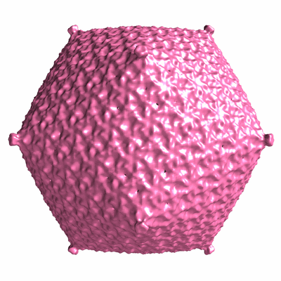
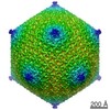
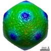
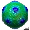
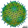
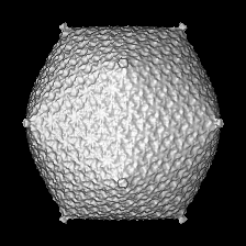
| Title | The structure of Sinorhizobium meliloti phage phiM12, a novel T=19 icosahedral phage that is the founder of a new group of T4-like phages | |||||||||
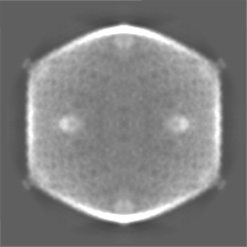
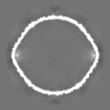
 Map data Map data | Cryo-EM reconstruction of phiM12, emptied of DNA | |||||||||
 Sample Sample |
| |||||||||
 Keywords Keywords | bacteriophage / phiM12 / Sinorhizobium meliloti / T=19 / rhizobia | |||||||||
| Biological species |  Sinorhizobium phage phiM12 (virus) Sinorhizobium phage phiM12 (virus) | |||||||||
| Method | single particle reconstruction / cryo EM / Resolution: 13.0 Å | |||||||||
 Authors Authors | Stroupe ME / Brewer TE / Sousa DR / Jones KM | |||||||||
 Citation Citation | Journal: Virology / Year: 2014 Title: The structure of Sinorhizobium meliloti phage ΦM12, which has a novel T=19l triangulation number and is the founder of a new group of T4-superfamily phages. Authors: M Elizabeth Stroupe / Tess E Brewer / Duncan R Sousa / Kathryn M Jones /  Abstract: ΦM12 is the first example of a T=19l geometry capsid, encapsulating the recently sequenced genome. Here, we present structures determined by cryo-EM of full and empty capsids. The structure reveals ...ΦM12 is the first example of a T=19l geometry capsid, encapsulating the recently sequenced genome. Here, we present structures determined by cryo-EM of full and empty capsids. The structure reveals the pattern for assembly of 1140 HK97-like capsid proteins, pointing to interactions at the pseudo 3-fold symmetry axes that hold together the asymmetric unit. The particular smooth surface of the capsid, along with a lack of accessory coat proteins encoded by the genome, suggest that this interface is the primary mechanism for capsid assembly. Two-dimensional averages of the tail, including the neck and baseplate, reveal that ΦM12 has a relatively narrow neck that attaches the tail to the capsid, as well as a three-layer baseplate. When free from DNA, the icosahedral edges expand by about 5nm, while the vertices stay at the same position, forming a similarly smooth, but bowed, T=19l icosahedral capsid. | |||||||||
| History |
|
- Structure visualization
| Movie |
 Movie viewer Movie viewer |
|---|---|
| Structure viewer | EM map: SurfViewMolmilJmol/JSmol |
| Supplemental images |
- Downloads & links
-EMDB archive
| Map data | emd_5718.map.gz | 19.1 MB | EMDB map data format | |
|---|---|---|---|---|
| Header (meta data) | emd-5718-v30.xmlemd-5718.xml | 9.3 KB 9.3 KB | Display Display | EMDB header |
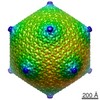
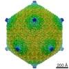
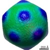
| Images |  400_5718.gif 400_5718.gif 80_5718.gif 80_5718.gif | 79.2 KB 5.2 KB | ||
| Archive directory |  http://ftp.pdbj.org/pub/emdb/structures/EMD-5718ftp://ftp.pdbj.org/pub/emdb/structures/EMD-5718 http://ftp.pdbj.org/pub/emdb/structures/EMD-5718ftp://ftp.pdbj.org/pub/emdb/structures/EMD-5718 | HTTPS FTP |
-Validation report
| Summary document | emd_5718_validation.pdf.gz | 78.7 KB | Display | EMDB validaton report |
|---|---|---|---|---|
| Full document | emd_5718_full_validation.pdf.gz | 77.8 KB | Display | |
| Data in XML | emd_5718_validation.xml.gz | 494 B | Display | |
| Arichive directory | https://ftp.pdbj.org/pub/emdb/validation_reports/EMD-5718ftp://ftp.pdbj.org/pub/emdb/validation_reports/EMD-5718 | HTTPS FTP |
-Related structure data

-Links
| EMDB pages | EMDB (EBI/PDBe) / EMDataResource |
|---|
-Map
| File | Download / File: emd_5718.map.gz / Format: CCP4 / Size: 41.9 MB / Type: IMAGE STORED AS FLOATING POINT NUMBER (4 BYTES) | ||||||||||||||||||||||||||||||||||||||||||||||||||||||||||||||||||||
|---|---|---|---|---|---|---|---|---|---|---|---|---|---|---|---|---|---|---|---|---|---|---|---|---|---|---|---|---|---|---|---|---|---|---|---|---|---|---|---|---|---|---|---|---|---|---|---|---|---|---|---|---|---|---|---|---|---|---|---|---|---|---|---|---|---|---|---|---|---|
| Annotation | Cryo-EM reconstruction of phiM12, emptied of DNA | ||||||||||||||||||||||||||||||||||||||||||||||||||||||||||||||||||||
| Projections & slices | Image control
Images are generated by Spider. | ||||||||||||||||||||||||||||||||||||||||||||||||||||||||||||||||||||
| Voxel size | X=Y=Z: 5.4 Å | ||||||||||||||||||||||||||||||||||||||||||||||||||||||||||||||||||||
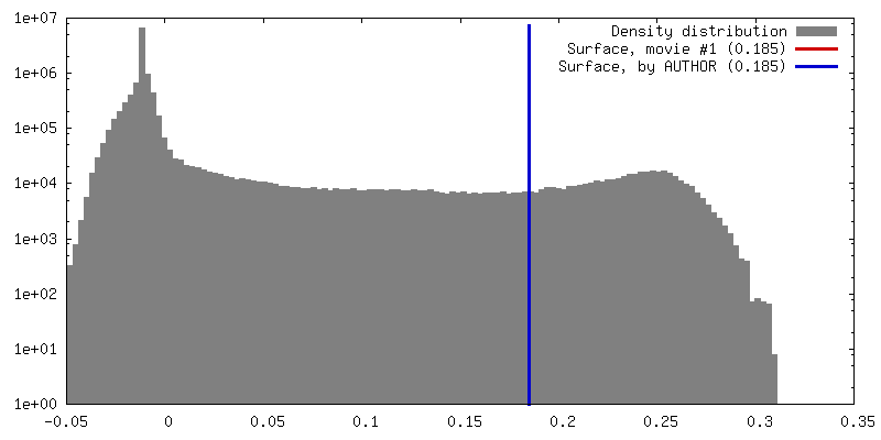
| Density |
| ||||||||||||||||||||||||||||||||||||||||||||||||||||||||||||||||||||
| Symmetry | Space group: 1 | ||||||||||||||||||||||||||||||||||||||||||||||||||||||||||||||||||||
| Details | EMDB XML:
CCP4 map header:
| ||||||||||||||||||||||||||||||||||||||||||||||||||||||||||||||||||||
 Z (Sec.)
 Y (Row.)
 X (Col.)


-Supplemental data
- Sample components
-Entire : Empty T=19 icosahedral shell from phiM12, a bacteriophage of Sino...
| Entire | Name: Empty T=19 icosahedral shell from phiM12, a bacteriophage of Sinorhizobium meliloti |
|---|---|
| Components |
|
-Supramolecule #1000: Empty T=19 icosahedral shell from phiM12, a bacteriophage of Sino...
| Supramolecule | Name: Empty T=19 icosahedral shell from phiM12, a bacteriophage of Sinorhizobium meliloti type: sample / ID: 1000 / Oligomeric state: icosahedral / Number unique components: 1 |
|---|
-Supramolecule #1: Sinorhizobium phage phiM12
| Supramolecule | Name: Sinorhizobium phage phiM12 / type: virus / ID: 1 / NCBI-ID: 1357423 / Sci species name: Sinorhizobium phage phiM12 / Sci species strain: phiM12 / Database: NCBI / Virus type: VIRION / Virus isolate: STRAIN / Virus enveloped: No / Virus empty: No |
|---|---|
| Host (natural) | Organism:  Sinorhizobium meliloti (bacteria) / Strain: 1021 / synonym: BACTERIA(EUBACTERIA) Sinorhizobium meliloti (bacteria) / Strain: 1021 / synonym: BACTERIA(EUBACTERIA) |
| Virus shell | Shell ID: 1 / Name: 1 / Diameter: 1000 Å / T number (triangulation number): 19 |
-Experimental details
-Structure determination
| Method | cryo EM |
|---|---|
 Processing Processing | single particle reconstruction |
| Aggregation state | particle |
-Sample preparation
| Buffer | pH: 7 / Details: 10 mM Na2HPO4, 1 mM MgSO4 |
|---|---|
| Grid | Details: 200 mesh 2/2 Quantifoil |
| Vitrification | Cryogen name: ETHANE / Chamber humidity: 100 % / Chamber temperature: 120 K / Instrument: FEI VITROBOT MARK IV / Method: blot 4 seconds |
- Electron microscopy
| Microscope | FEI TITAN KRIOS |
|---|---|
| Date | May 13, 2013 |
| Image recording | Category: CCD / Film or detector model: GATAN ULTRASCAN 4000 (4k x 4k) / Number real images: 1000 / Average electron dose: 15 e/Å2 |
| Tilt angle min | 0 |
| Tilt angle max | 0 |
| Electron beam | Acceleration voltage: 120 kV / Electron source:  FIELD EMISSION GUN FIELD EMISSION GUN |
| Electron optics | Calibrated magnification: 65555 / Illumination mode: FLOOD BEAM / Imaging mode: BRIGHT FIELD / Nominal defocus max: 3.5 µm / Nominal defocus min: 1.5 µm / Nominal magnification: 59000 |
| Sample stage | Specimen holder model: FEI TITAN KRIOS AUTOGRID HOLDER |
| Experimental equipment |  Model: Titan Krios / Image courtesy: FEI Company |
-Image processing
| Details | Images were selected by hand; initial model determined in EMAN and refined in EMAN and Frealign |
|---|---|
| CTF correction | Details: micrograph |
| Final reconstruction | Algorithm: OTHER / Resolution.type: BY AUTHOR / Resolution: 13.0 Å / Resolution method: FSC 0.143 CUT-OFF / Software - Name: EMAN, FREALIGN / Number images used: 2038 |
