 Movie
Movie Controller


+ Open data

- Basic information
| Entry |  | ||||||||||||
|---|---|---|---|---|---|---|---|---|---|---|---|---|---|
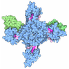
| Title | Cryo-EM structure of the human LENG8-PCID2-SEM1 complex | ||||||||||||
 Map data Map data | Cryo-EM map of the human LENG8-PCID2-SEM1 complex (Map D) | ||||||||||||
 Sample Sample |
| ||||||||||||
 Keywords Keywords | mRNA export / GENE REGULATION | ||||||||||||
| Function / homology |  Function and homology information Function and homology informationnegative regulation of lymphoid progenitor cell differentiation / post-transcriptional tethering of RNA polymerase II gene DNA at nuclear periphery / nuclear pore nuclear basket / spleen development / Impaired BRCA2 translocation to the nucleus / Impaired BRCA2 binding to SEM1 (DSS1) / integrator complex / transcription export complex 2 / proteasome regulatory particle, lid subcomplex / positive regulation of B cell differentiation ...negative regulation of lymphoid progenitor cell differentiation / post-transcriptional tethering of RNA polymerase II gene DNA at nuclear periphery / nuclear pore nuclear basket / spleen development / Impaired BRCA2 translocation to the nucleus / Impaired BRCA2 binding to SEM1 (DSS1) / integrator complex / transcription export complex 2 / proteasome regulatory particle, lid subcomplex / positive regulation of B cell differentiation / poly(A)+ mRNA export from nucleus / Regulation of ornithine decarboxylase (ODC) / Proteasome assembly / cellular response to type I interferon / Cross-presentation of soluble exogenous antigens (endosomes) / negative regulation of gene expression, epigenetic / Somitogenesis / Homologous DNA Pairing and Strand Exchange / Defective homologous recombination repair (HRR) due to BRCA1 loss of function / Defective HDR through Homologous Recombination Repair (HRR) due to PALB2 loss of BRCA1 binding function / Defective HDR through Homologous Recombination Repair (HRR) due to PALB2 loss of BRCA2/RAD51/RAD51C binding function / Resolution of D-loop Structures through Synthesis-Dependent Strand Annealing (SDSA) / Resolution of D-loop Structures through Holliday Junction Intermediates / detection of maltose stimulus / carbohydrate transport / Impaired BRCA2 binding to RAD51 / carbohydrate transmembrane transporter activity / Presynaptic phase of homologous DNA pairing and strand exchange / maltose binding / maltose transport / maltodextrin transmembrane transport / proteasome assembly / mRNA export from nucleus / ATP-binding cassette (ABC) transporter complex, substrate-binding subunit-containing / proteasome complex / ATP-binding cassette (ABC) transporter complex / Activation of STAT3 by cadherin engagement / regulation of proteasomal protein catabolic process / cell chemotaxis / ubiquitin binding / Regulation of activated PAK-2p34 by proteasome mediated degradation / proteasomal protein catabolic process / Autodegradation of Cdh1 by Cdh1:APC/C / APC/C:Cdc20 mediated degradation of Securin / Asymmetric localization of PCP proteins / Ubiquitin-dependent degradation of Cyclin D / SCF-beta-TrCP mediated degradation of Emi1 / AUF1 (hnRNP D0) binds and destabilizes mRNA / NIK-->noncanonical NF-kB signaling / TNFR2 non-canonical NF-kB pathway / Assembly of the pre-replicative complex / Vpu mediated degradation of CD4 / Cdc20:Phospho-APC/C mediated degradation of Cyclin A / Dectin-1 mediated noncanonical NF-kB signaling / Degradation of DVL / Degradation of AXIN / Degradation of CRY and PER proteins / transcription elongation by RNA polymerase II / Hh mutants are degraded by ERAD / Activation of NF-kappaB in B cells / G2/M Checkpoints / Degradation of GLI1 by the proteasome / Hedgehog ligand biogenesis / Regulation of RUNX3 expression and activity / Autodegradation of the E3 ubiquitin ligase COP1 / Defective CFTR causes cystic fibrosis / GSK3B and BTRC:CUL1-mediated-degradation of NFE2L2 / Negative regulation of NOTCH4 signaling / AMPK-induced ERAD and lysosome mediated degradation of PD-L1(CD274) / Hedgehog 'on' state / APC/C:Cdh1 mediated degradation of Cdc20 and other APC/C:Cdh1 targeted proteins in late mitosis/early G1 / Vif-mediated degradation of APOBEC3G / FBXL7 down-regulates AURKA during mitotic entry and in early mitosis / Degradation of GLI2 by the proteasome / GLI3 is processed to GLI3R by the proteasome / MAPK6/MAPK4 signaling / GSK3B-mediated proteasomal degradation of PD-L1(CD274) / Degradation of CDH1 / Degradation of beta-catenin by the destruction complex / Oxygen-dependent proline hydroxylation of Hypoxia-inducible Factor Alpha / HDR through Homologous Recombination (HRR) / CDK-mediated phosphorylation and removal of Cdc6 / ABC-family protein mediated transport / CLEC7A (Dectin-1) signaling / SCF(Skp2)-mediated degradation of p27/p21 / FCERI mediated NF-kB activation / SPOP-mediated proteasomal degradation of PD-L1(CD274) / Regulation of expression of SLITs and ROBOs / Regulation of PTEN stability and activity / Ubiquitin-Mediated Degradation of Phosphorylated Cdc25A / Ribosome Quality Control (RQC) complex extracts and degrades nascent peptide / Interleukin-1 signaling / Orc1 removal from chromatin / Regulation of RUNX2 expression and activity / Regulation of RAS by GAPs / The role of GTSE1 in G2/M progression after G2 checkpoint / Separation of Sister Chromatids / KEAP1-NFE2L2 pathway / UCH proteinases / Downstream TCR signaling Similarity search - Function | ||||||||||||
| Biological species |  Homo sapiens (human) Homo sapiens (human) | ||||||||||||
| Method | single particle reconstruction / cryo EM / Resolution: 3.5 Å | ||||||||||||
 Authors Authors | Hohmann U / Graf M / Plaschka C | ||||||||||||
| Funding support | European Union, 3 items
| ||||||||||||
 Citation Citation | Journal: Nature / Year: 2026 Title: Molecular basis of polyadenylated RNA fate determination in the nucleus. Authors: Andrii Bugai / Ulrich Hohmann / Ana Lorenzo / Max Graf / Laura Fin / Jérôme O Rouvière / Laszlo Tirian / Yuhui Dou / Marion Le Rest / Patrik Polák / Dennis Johnsen / Lis Jakobsen / Jens ...Authors: Andrii Bugai / Ulrich Hohmann / Ana Lorenzo / Max Graf / Laura Fin / Jérôme O Rouvière / Laszlo Tirian / Yuhui Dou / Marion Le Rest / Patrik Polák / Dennis Johnsen / Lis Jakobsen / Jens Skorstengaard Andersen / Julius Brennecke / Clemens Plaschka / Torben Heick Jensen /     Abstract: Eukaryotic genomes generate a plethora of polyadenylated (pA) RNAs, which are packaged into ribonucleoprotein particles (RNPs). To ensure faithful gene expression, functional pA RNPs, including ...Eukaryotic genomes generate a plethora of polyadenylated (pA) RNAs, which are packaged into ribonucleoprotein particles (RNPs). To ensure faithful gene expression, functional pA RNPs, including protein-coding RNPs, are exported to the cytoplasm, whereas transcripts within non-functional pA RNPs are degraded in the nucleus. How cells distinguish these opposing fates remains unknown. The DExD-box ATPase UAP56 (also known as DDX39B) is a central component of functional pA RNPs, and promotes their docking to the nuclear pore complex-anchored TREX-2, which triggers transcript release from UAP56 to facilitate export. Here we reveal that the poly(A) tail exosome targeting (PAXT) connection binds a TREX-2-like module, which releases pA RNAs from UAP56 for decay by the nuclear exosome. The core of this module consists of a LENG8-PCID2-SEM1 trimer, which we show is structurally and biochemically equivalent to the central GANP-PCID2-SEM1 trimer of TREX-2. Mutagenesis and transcriptomic data demonstrate that the nuclear fate of pA RNPs is governed by the contending actions of nucleoplasmic PAXT and nuclear pore complex-associated TREX-2, which interpret RNA-bound UAP56 as a signal for RNA decay or export, respectively. As RNA targets of PAXT are generally short and intron-poor, we propose an overall model for pA RNP fate determination whereby the distinct sub-nuclear localizations of PAXT and TREX-2 govern the degradation of short non-functional pA RNAs while allowing export of their longer and functional counterparts. | ||||||||||||
| History |
|
- Structure visualization
| Supplemental images |
|---|

- Downloads & links
-EMDB archive
| Map data | emd_56931.map.gz | 59.6 MB | EMDB map data format | |
|---|---|---|---|---|
| Header (meta data) | emd-56931-v30.xmlemd-56931.xml | 23.5 KB 23.5 KB | Display Display | EMDB header |
| Images |  emd_56931.png emd_56931.png | 132.9 KB | ||
| Filedesc metadata | emd-56931.cif.gz | 6.9 KB | ||
| Others | emd_56931_half_map_1.map.gzemd_56931_half_map_2.map.gz | 59.3 MB 59.3 MB | ||
| Archive directory |  https://data.pdbj.org/pub/emdb/structures/EMD-56931ftp://data.pdbj.org/pub/emdb/structures/EMD-56931 https://data.pdbj.org/pub/emdb/structures/EMD-56931ftp://data.pdbj.org/pub/emdb/structures/EMD-56931 | HTTPS FTP |
-Related structure data
| Related structure data |  28wzMC  28wyC  28xaC  28xbC  9rv1C M: atomic model generated by this map C: citing same article ( |
|---|---|
| Similar structure data |


-Links
| EMDB pages | EMDB (EBI/PDBe) / EMDataResource |
|---|---|
| Related items in Molecule of the Month |







-Map
| File | Download / File: emd_56931.map.gz / Format: CCP4 / Size: 64 MB / Type: IMAGE STORED AS FLOATING POINT NUMBER (4 BYTES) | ||||||||||||||||||||||||||||||||||||
|---|---|---|---|---|---|---|---|---|---|---|---|---|---|---|---|---|---|---|---|---|---|---|---|---|---|---|---|---|---|---|---|---|---|---|---|---|---|
| Annotation | Cryo-EM map of the human LENG8-PCID2-SEM1 complex (Map D) | ||||||||||||||||||||||||||||||||||||
| Projections & slices | Image control
Images are generated by Spider. | ||||||||||||||||||||||||||||||||||||
| Voxel size | X=Y=Z: 0.878 Å | ||||||||||||||||||||||||||||||||||||
| Density |
| ||||||||||||||||||||||||||||||||||||
| Symmetry | Space group: 1 | ||||||||||||||||||||||||||||||||||||
| Details | EMDB XML:
|
 Z (Sec.)
 Y (Row.)
 X (Col.)


-Supplemental data
-Half map: Cryo-EM map of the human LENG8-PCID2-SEM1 complex (Map D), half map A
| File | emd_56931_half_map_1.map | ||||||||||||
|---|---|---|---|---|---|---|---|---|---|---|---|---|---|
| Annotation | Cryo-EM map of the human LENG8-PCID2-SEM1 complex (Map D), half map A | ||||||||||||
| Projections & Slices |
| ||||||||||||
| Density Histograms |


-Half map: Cryo-EM map of the human LENG8-PCID2-SEM1 complex (Map D), half map B
| File | emd_56931_half_map_2.map | ||||||||||||
|---|---|---|---|---|---|---|---|---|---|---|---|---|---|
| Annotation | Cryo-EM map of the human LENG8-PCID2-SEM1 complex (Map D), half map B | ||||||||||||
| Projections & Slices |
| ||||||||||||
| Density Histograms |


- Sample components
-Entire : Human LENG8-PCID2-SEM1 complex
| Entire | Name: Human LENG8-PCID2-SEM1 complex |
|---|---|
| Components |
|
-Supramolecule #1: Human LENG8-PCID2-SEM1 complex
| Supramolecule | Name: Human LENG8-PCID2-SEM1 complex / type: complex / ID: 1 / Parent: 0 / Macromolecule list: all |
|---|---|
| Source (natural) | Organism: Homo sapiens (human) |
-Supramolecule #2: Human LENG8-PCID2-SEM1 complex
| Supramolecule | Name: Human LENG8-PCID2-SEM1 complex / type: complex / ID: 2 / Parent: 1 / Macromolecule list: all |
|---|---|
| Source (natural) | Organism: Homo sapiens (human) |
-Macromolecule #1: Maltose/maltodextrin-binding periplasmic protein,Leukocyte recept...
| Macromolecule | Name: Maltose/maltodextrin-binding periplasmic protein,Leukocyte receptor cluster member 8 type: protein_or_peptide / ID: 1 / Number of copies: 1 / Enantiomer: LEVO |
|---|---|
| Source (natural) | Organism: Homo sapiens (human) |
| Molecular weight | Theoretical: 76.698383 KDa |
| Recombinant expression | Organism:   Escherichia coli (E. coli) Escherichia coli (E. coli) |
| Sequence | String: KIEEGKLVIW INGDKGYNGL AEVGKKFEKD TGIKVTVEHP DKLEEKFPQV AATGDGPDII FWAHDRFGGY AQSGLLAEIT PDKAFQDKL YPFTWDAVRY NGKLIAYPIA VEALSLIYNK DLLPNPPKTW EEIPALDKEL KAKGKSALMF NLQEPYFTWP L IAADGGYA ...String: KIEEGKLVIW INGDKGYNGL AEVGKKFEKD TGIKVTVEHP DKLEEKFPQV AATGDGPDII FWAHDRFGGY AQSGLLAEIT PDKAFQDKL YPFTWDAVRY NGKLIAYPIA VEALSLIYNK DLLPNPPKTW EEIPALDKEL KAKGKSALMF NLQEPYFTWP L IAADGGYA FKYENGKYDI KDVGVDNAGA KAGLTFLVDL IKNKHMNADT DYSIAEAAFN KGETAMTING PWAWSNIDTS KV NYGVTVL PTFKGQPSKP FVGVLSAGIN AASPNKELAK EFLENYLLTD EGLEAVNKDK PLGAVALKSY EEELAKDPRI AAT MENAQK GEIMPNIPQM SAFWYAVRTA VINAASGRQT VDEALKDAQT SSGLEVLFQG PSRKKMAALE CEDPERELKK QKRA ARFQH GHSRRLRLEP LVLQMSSLES SGADPDWQEL QIVGTCPDIT KHYLRLTCAP DPSTVRPVAV LKKSLCMVKC HWKEK QDYA FACEQMKSIR QDLTVQGIRT EFTVEVYETH ARIALEKGDH EEFNQCQTQL KSLYAENLPG NVGEFTAYRI LYYIFT KNS GDITTELAYL TRELKADPCV AHALALRTAW ALGNYHRFFR LYCHAPCMSG YLVDKFADRE RKVALKAMIK TFRPALP VS YLQAELAFEG EAACRAFLEP LGLAYTGPDN SSIDCRLSLA QLSAF UniProtKB: Maltose/maltodextrin-binding periplasmic protein, Leukocyte receptor cluster member 8 |
-Macromolecule #2: PCI domain-containing protein 2
| Macromolecule | Name: PCI domain-containing protein 2 / type: protein_or_peptide / ID: 2 / Number of copies: 1 / Enantiomer: LEVO |
|---|---|
| Source (natural) | Organism: Homo sapiens (human) |
| Molecular weight | Theoretical: 51.713254 KDa |
| Recombinant expression | Organism: Escherichia coli (E. coli) |
| Sequence | String: GKPIPNPLLG LDSTGSGKPI PNPLLGLDST GSGKPIPNPL LGLDSTSSGL EVLFQGPMAH ITINQYLQQV YEAIDSRDGA SCAELVSFK HPHVANPRLQ MASPEEKCQQ VLEPPYDEMF AAHLRCTYAV GNHDFIEAYK CQTVIVQSFL RAFQAHKEEN W ALPVMYAV ...String: GKPIPNPLLG LDSTGSGKPI PNPLLGLDST GSGKPIPNPL LGLDSTSSGL EVLFQGPMAH ITINQYLQQV YEAIDSRDGA SCAELVSFK HPHVANPRLQ MASPEEKCQQ VLEPPYDEMF AAHLRCTYAV GNHDFIEAYK CQTVIVQSFL RAFQAHKEEN W ALPVMYAV ALDLRVFANN ADQQLVKKGK SKVGDMLEKA AELLMSCFRV CASDTRAGIE DSKKWGMLFL VNQLFKIYFK IN KLHLCKP LIRAIDSSNL KDDYSTAQRV TYKYYVGRKA MFDSDFKQAE EYLSFAFEHC HRSSQKNKRM ILIYLLPVKM LLG HMPTVE LLKKYHLMQF AEVTRAVSEG NLLLLHEALA KHEAFFIRCG IFLILEKLKI ITYRNLFKKV YLLLKTHQLS LDAF LVALK FMQVEDVDID EVQCILANLI YMGHVKGYIS HQHQKLVVSK QNPFPPLSTV C UniProtKB: PCI domain-containing protein 2 |
-Macromolecule #3: 26S proteasome complex subunit SEM1
| Macromolecule | Name: 26S proteasome complex subunit SEM1 / type: protein_or_peptide / ID: 3 / Number of copies: 1 / Enantiomer: LEVO |
|---|---|
| Source (natural) | Organism: Homo sapiens (human) |
| Molecular weight | Theoretical: 8.284611 KDa |
| Recombinant expression | Organism: Escherichia coli (E. coli) |
| Sequence | String: MSEKKQPVDL GLLEEDDEFE EFPAEDWAGL DEDEDAHVWE DNWDDDNVED DFSNQLRAEL EKHGYKMETS UniProtKB: 26S proteasome complex subunit SEM1 |
-Experimental details
-Structure determination
| Method | cryo EM |
|---|---|
 Processing Processing | single particle reconstruction |
| Aggregation state | particle |
-Sample preparation
| Concentration | 0.5 mg/mL |
|---|---|
| Buffer | pH: 7.9 |
| Vitrification | Cryogen name: ETHANE / Chamber humidity: 95 % / Chamber temperature: 281 K / Instrument: LEICA EM GP |
- Electron microscopy
| Microscope | TFS KRIOS |
|---|---|
| Image recording | Film or detector model: FEI FALCON IV (4k x 4k) / Average electron dose: 50.0 e/Å2 |
| Electron beam | Acceleration voltage: 300 kV / Electron source:  FIELD EMISSION GUN FIELD EMISSION GUN |
| Electron optics | Illumination mode: OTHER / Imaging mode: BRIGHT FIELD / Nominal defocus max: 2.5 µm / Nominal defocus min: 1.0 µm |
| Sample stage | Cooling holder cryogen: NITROGEN |
| Experimental equipment |  Model: Titan Krios / Image courtesy: FEI Company |
