 Movie
Movie Controller

[English] 日本語
 Yorodumi
- EMDB-5476: Structure of the vacuolar-type ATPase from Saccharomyces cerevisi... -
+ Open data

- Basic information
| Entry | Database: EMDB / ID: EMD-5476 | |||||||||
|---|---|---|---|---|---|---|---|---|---|---|
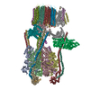
| Title | Structure of the vacuolar-type ATPase from Saccharomyces cerevisiae at 11 Angstrom resolution | |||||||||
 Map data Map data | 3D map of V-type ATPase | |||||||||
 Sample Sample |
| |||||||||
 Keywords Keywords | membrane protein / proton pump / ATPase / vacuole / endosome / lysosome / plasma membrane / Golgi | |||||||||
| Biological species |  | |||||||||
| Method | single particle reconstruction / cryo EM / Resolution: 11.0 Å | |||||||||
 Authors Authors | Benlekbir S / Bueler SA / Rubinstein JL | |||||||||
 Citation Citation | Journal: Nat Struct Mol Biol / Year: 2012 Title: Structure of the vacuolar-type ATPase from Saccharomyces cerevisiae at 11-Å resolution. Authors: Samir Benlekbir / Stephanie A Bueler / John L Rubinstein /  Abstract: Vacuolar-type ATPases (V-type ATPases) in eukaryotic cells are large membrane protein complexes that acidify various intracellular compartments. The enzymes are regulated by dissociation of the V(1) ...Vacuolar-type ATPases (V-type ATPases) in eukaryotic cells are large membrane protein complexes that acidify various intracellular compartments. The enzymes are regulated by dissociation of the V(1) and V(O) regions of the complex. Here we present the structure of the Saccharomyces cerevisiae V-type ATPase at 11-Å resolution by cryo-EM of protein particles in ice. The structure explains many cross-linking and protein interaction studies. Docking of crystal structures suggests that inhibition of ATPase activity by the dissociated V(1) region involves rearrangement of the N- and C-terminal domains of subunit H and also suggests how this inhibition is triggered upon dissociation. We provide support for this model by demonstrating that mutation of subunit H to increase the rigidity of the linker between its two domains decreases its ability to inhibit ATPase activity. | |||||||||
| History |
|
- Structure visualization
| Movie |
 Movie viewer Movie viewer |
|---|---|
| Structure viewer | EM map: SurfViewMolmilJmol/JSmol |
| Supplemental images |

- Downloads & links
-EMDB archive
| Map data | emd_5476.map.gz | 31.4 MB | EMDB map data format | |
|---|---|---|---|---|
| Header (meta data) | emd-5476-v30.xmlemd-5476.xml | 50.3 KB 50.3 KB | Display Display | EMDB header |
| Images | 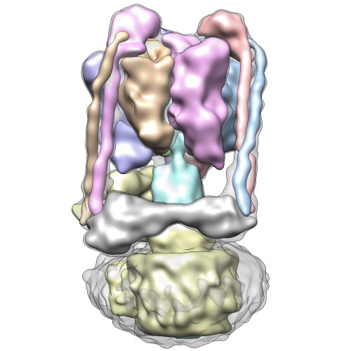 emd_5476_1.jpg emd_5476_1.jpg | 43.4 KB | ||
| Masks | emd_5476_msk_1.mapemd_5476_msk_10.mapemd_5476_msk_11.mapemd_5476_msk_12.mapemd_5476_msk_13.mapemd_5476_msk_14.mapemd_5476_msk_15.mapemd_5476_msk_16.mapemd_5476_msk_17.mapemd_5476_msk_18.mapemd_5476_msk_19.mapemd_5476_msk_2.mapemd_5476_msk_3.mapemd_5476_msk_4.mapemd_5476_msk_5.mapemd_5476_msk_6.mapemd_5476_msk_7.mapemd_5476_msk_8.mapemd_5476_msk_9.map | 64 MB 64 MB 64 MB 64 MB 64 MB 64 MB 64 MB 64 MB 64 MB 64 MB 64 MB 64 MB 64 MB 64 MB 64 MB 64 MB 64 MB 64 MB 64 MB | Mask map | |
| Archive directory |  http://ftp.pdbj.org/pub/emdb/structures/EMD-5476ftp://ftp.pdbj.org/pub/emdb/structures/EMD-5476 http://ftp.pdbj.org/pub/emdb/structures/EMD-5476ftp://ftp.pdbj.org/pub/emdb/structures/EMD-5476 | HTTPS FTP |
-Related structure data
| Similar structure data |
|---|


-Links
| EMDB pages | EMDB (EBI/PDBe) / EMDataResource |
|---|
-Map
| File | Download / File: emd_5476.map.gz / Format: CCP4 / Size: 62.5 MB / Type: IMAGE STORED AS FLOATING POINT NUMBER (4 BYTES) | ||||||||||||||||||||||||||||||||||||||||||||||||||||||||||||||||||||
|---|---|---|---|---|---|---|---|---|---|---|---|---|---|---|---|---|---|---|---|---|---|---|---|---|---|---|---|---|---|---|---|---|---|---|---|---|---|---|---|---|---|---|---|---|---|---|---|---|---|---|---|---|---|---|---|---|---|---|---|---|---|---|---|---|---|---|---|---|---|
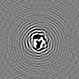
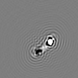
| Annotation | 3D map of V-type ATPase | ||||||||||||||||||||||||||||||||||||||||||||||||||||||||||||||||||||
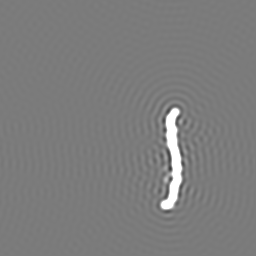
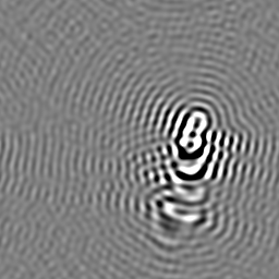
| Projections & slices | Image control
Images are generated by Spider. | ||||||||||||||||||||||||||||||||||||||||||||||||||||||||||||||||||||
| Voxel size | X=Y=Z: 1.4 Å | ||||||||||||||||||||||||||||||||||||||||||||||||||||||||||||||||||||
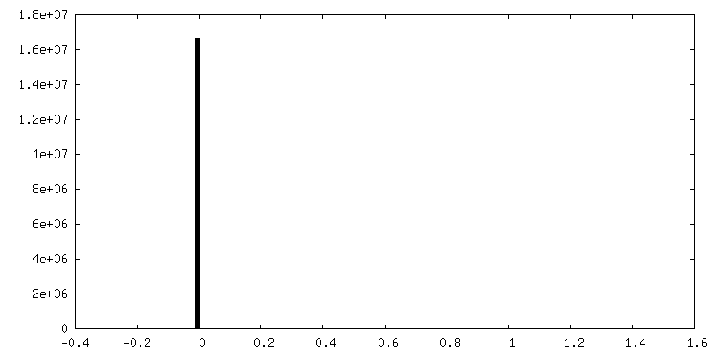
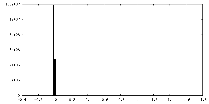
| Density |
| ||||||||||||||||||||||||||||||||||||||||||||||||||||||||||||||||||||
| Symmetry | Space group: 1 | ||||||||||||||||||||||||||||||||||||||||||||||||||||||||||||||||||||
| Details | EMDB XML:
CCP4 map header:
| ||||||||||||||||||||||||||||||||||||||||||||||||||||||||||||||||||||
 Z (Sec.)
 Y (Row.)
 X (Col.)


-Supplemental data
+Segmentation: A subunit 2


+Segmentation: E subunit 3


+Segmentation: G subunit 1


+Segmentation: G subunit 2


+Segmentation: G subunit 3


+Segmentation: H subunit


+Segmentation: a subunit


+Segmentation: c-ring


+Segmentation: d subunit


+Segmentation: detergent/lipid plug


+Segmentation: A subunit 1


+Segmentation: A subunit 3


+Segmentation: B subunit 1


+Segmentation: B subunit 2


+Segmentation: B subunit 3


+Segmentation: C subunit


+Segmentation: DF subcomplex


+Segmentation: E subunit 1


+Segmentation: E subunit 2


- Sample components
-Entire : vacuolar-type ATPases
| Entire | Name: vacuolar-type ATPases |
|---|---|
| Components |
|
-Supramolecule #1000: vacuolar-type ATPases
| Supramolecule | Name: vacuolar-type ATPases / type: sample / ID: 1000 Oligomeric state: A3B3CDE3FG3HadcXc'Yc''Z complex where X, Y, and Z indicate unknown stoichiometry Number unique components: 1 |
|---|---|
| Molecular weight | Experimental: 900 KDa / Theoretical: 900 KDa / Method: Gel filtration |
-Macromolecule #1: vacuolar-type ATPases
| Macromolecule | Name: vacuolar-type ATPases / type: protein_or_peptide / ID: 1 / Name.synonym: V-ATPase / Details: Detergent solubilized protein complex / Number of copies: 1 / Oligomeric state: monomer / Recombinant expression: No / Database: NCBI |
|---|---|
| Source (natural) | Organism: |
| Molecular weight | Experimental: 900 KDa / Theoretical: 900 KDa |
-Experimental details
-Structure determination
| Method | cryo EM |
|---|---|
 Processing Processing | single particle reconstruction |
| Aggregation state | particle |
-Sample preparation
| Concentration | 2.5 mg/mL |
|---|---|
| Buffer | pH: 7.4 Details: 50 mM Tris-HCl, 150 mM NaCl, 0.03% w/v dodecylmaltoside |
| Grid | Details: Quantifoil R2/2 glow discharged in air |
| Vitrification | Cryogen name: ETHANE / Chamber humidity: 100 % / Instrument: FEI VITROBOT MARK III / Method: Blot for 20 seconds before freezing |
- Electron microscopy
| Microscope | FEI TECNAI F20 |
|---|---|
| Alignment procedure | Legacy - Astigmatism: Manually corrected by inspecting FFT |
| Date | Jan 1, 2011 |
| Image recording | Category: FILM / Film or detector model: KODAK SO-163 FILM / Digitization - Scanner: ZEISS SCAI / Digitization - Sampling interval: 7 µm / Number real images: 1000 / Average electron dose: 12 e/Å2 / Bits/pixel: 8 |
| Electron beam | Acceleration voltage: 200 kV / Electron source:  FIELD EMISSION GUN FIELD EMISSION GUN |
| Electron optics | Calibrated magnification: 50000 / Illumination mode: FLOOD BEAM / Imaging mode: BRIGHT FIELD / Cs: 2.0 mm / Nominal defocus max: 5.0 µm / Nominal defocus min: 3.0 µm / Nominal magnification: 50000 |
| Sample stage | Specimen holder model: GATAN LIQUID NITROGEN |
| Experimental equipment |  Model: Tecnai F20 / Image courtesy: FEI Company |
-Image processing
| Details | particles selected manually with Ximdisp |
|---|---|
| CTF correction | Details: Each particle |
| Final reconstruction | Algorithm: OTHER / Resolution.type: BY AUTHOR / Resolution: 11.0 Å / Resolution method: OTHER / Software - Name: Search_Fspace, Refine_Fspace, Build_Fspace / Number images used: 34448 |
