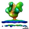
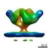
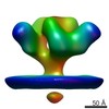
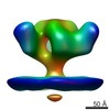
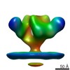
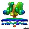
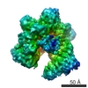
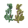
ジャーナル: PLoS Pathog / 年: 2012 タイトル: Structural mechanism of trimeric HIV-1 envelope glycoprotein activation. 著者: Erin E H Tran / Mario J Borgnia / Oleg Kuybeda / David M Schauder / Alberto Bartesaghi / Gabriel A Frank / Guillermo Sapiro / Jacqueline L S Milne / Sriram Subramaniam / 要旨: HIV-1 infection begins with the binding of trimeric viral envelope glycoproteins (Env) to CD4 and a co-receptor on target T-cells. Understanding how these ligands influence the structure of Env is of ...HIV-1 infection begins with the binding of trimeric viral envelope glycoproteins (Env) to CD4 and a co-receptor on target T-cells. Understanding how these ligands influence the structure of Env is of fundamental interest for HIV vaccine development. Using cryo-electron microscopy, we describe the contrasting structural outcomes of trimeric Env binding to soluble CD4, to the broadly neutralizing, CD4-binding site antibodies VRC01, VRC03 and b12, or to the monoclonal antibody 17b, a co-receptor mimic. Binding of trimeric HIV-1 BaL Env to either soluble CD4 or 17b alone, is sufficient to trigger formation of the open quaternary conformation of Env. In contrast, VRC01 locks Env in the closed state, while b12 binding requires a partial opening in the quaternary structure of trimeric Env. Our results show that, despite general similarities in regions of the HIV-1 gp120 polypeptide that contact CD4, VRC01, VRC03 and b12, there are important differences in quaternary structures of the complexes these ligands form on native trimeric Env, and potentially explain differences in the neutralizing breadth and potency of antibodies with similar specificities. From cryo-electron microscopic analysis at ∼9 Å resolution of a cleaved, soluble version of trimeric Env, we show that a structural signature of the open Env conformation is a three-helix motif composed of α-helical segments derived from highly conserved, non-glycosylated N-terminal regions of the gp41 trimer. The three N-terminal gp41 helices in this novel, activated Env conformation are held apart by their interactions with the rest of Env, and are less compactly packed than in the post-fusion, six-helix bundle state. These findings suggest a new structural template for designing immunogens that can elicit antibodies targeting HIV at a vulnerable, pre-entry stage.
生物種: Homo sapiens (ヒト) / 組換プラスミド: SOSIP-PPI4 and furin-pcDNA3.1
-
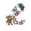
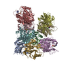
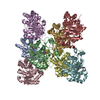
分子 #2: Fab portion of monoclonal antibody 17b
分子
名称: Fab portion of monoclonal antibody 17b / タイプ: protein_or_peptide / ID: 2 / Name.synonym: 17b Fab / 詳細: Fab fragment / 組換発現: No / データベース: NCBI
由来(天然)
生物種: unidentified (未定義)
分子量
理論値: 50 KDa
-
実験情報
-
構造解析
手法
クライオ電子顕微鏡法
解析
サブトモグラム平均法
試料の集合状態
particle
-
試料調製
濃度
0.42 mg/mL
緩衝液
pH: 7.5 / 詳細: TNE Buffer (10 mM Tris, 150 mM NaCl, 1 mM EDTA)
グリッド
詳細: Protochips C-flat R 2/2, plasma cleaned
凍結
凍結剤: ETHANE / チャンバー内湿度: 100 % / 装置: FEI VITROBOT MARK III 手法: blot for 6 seconds, blot offset of -2, plunge into an ethane slurry cooled by liquid nitrogen
 ムービー
ムービー コントローラー


 データを開く
 基本情報
基本情報 マップデータ
 試料
試料 キーワード
キーワード
 Human immunodeficiency virus 1 (ヒト免疫不全ウイルス) / unidentified (未定義)
 データ登録者
 引用
引用
 構造の表示
構造の表示 ムービービューア

 ダウンロードとリンク
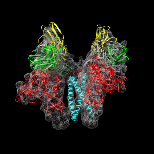 emd_5462.png
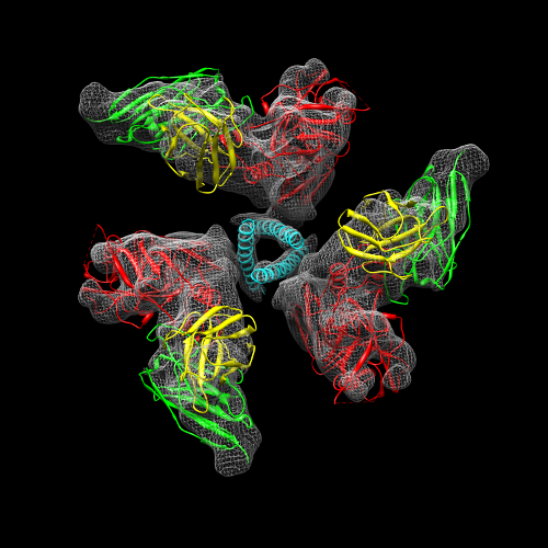 emd_5462_1.png
 http://ftp.pdbj.org/pub/emdb/structures/EMD-5462


 Z (Sec.)
 Y (Row.)
 X (Col.)


 試料の構成要素
 Homo sapiens (ヒト) / 組換プラスミド: SOSIP-PPI4 and furin-pcDNA3.1
 解析
解析 電子顕微鏡法
 FIELD EMISSION GUN