Movie
Movie Controller

[English] 日本語
 Yorodumi
- EMDB-5462: Cryo-electron tomography of a trimeric soluble HIV Env construct,... -
+ Open data

- Basic information
| Entry | Database: EMDB / ID: EMD-5462 | |||||||||
|---|---|---|---|---|---|---|---|---|---|---|
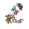
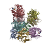
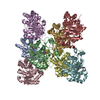
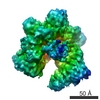
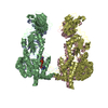
| Title | Cryo-electron tomography of a trimeric soluble HIV Env construct, gp140 SOSIP, in complex with Fab 17b | |||||||||
 Map data Map data | Reconstruction of soluble KNH1144-SOSIP Env trimers in complex with Fab 17b | |||||||||
 Sample Sample |
| |||||||||
 Keywords Keywords | HIV / gp140 / SOSIP / 17b | |||||||||
| Biological species |   Human immunodeficiency virus 1 / unidentified (others) Human immunodeficiency virus 1 / unidentified (others) | |||||||||
| Method | subtomogram averaging / cryo EM / Resolution: 8.8 Å | |||||||||
 Authors Authors | Tran EEH / Borgnia MJ / Kuybeda O / Schauder DM / Bartesaghi A / Frank GA / Sapiro G / Milne JLS / Subramaniam S | |||||||||
 Citation Citation | Journal: PLoS Pathog / Year: 2012 Title: Structural mechanism of trimeric HIV-1 envelope glycoprotein activation. Authors: Erin E H Tran / Mario J Borgnia / Oleg Kuybeda / David M Schauder / Alberto Bartesaghi / Gabriel A Frank / Guillermo Sapiro / Jacqueline L S Milne / Sriram Subramaniam /  Abstract: HIV-1 infection begins with the binding of trimeric viral envelope glycoproteins (Env) to CD4 and a co-receptor on target T-cells. Understanding how these ligands influence the structure of Env is of ...HIV-1 infection begins with the binding of trimeric viral envelope glycoproteins (Env) to CD4 and a co-receptor on target T-cells. Understanding how these ligands influence the structure of Env is of fundamental interest for HIV vaccine development. Using cryo-electron microscopy, we describe the contrasting structural outcomes of trimeric Env binding to soluble CD4, to the broadly neutralizing, CD4-binding site antibodies VRC01, VRC03 and b12, or to the monoclonal antibody 17b, a co-receptor mimic. Binding of trimeric HIV-1 BaL Env to either soluble CD4 or 17b alone, is sufficient to trigger formation of the open quaternary conformation of Env. In contrast, VRC01 locks Env in the closed state, while b12 binding requires a partial opening in the quaternary structure of trimeric Env. Our results show that, despite general similarities in regions of the HIV-1 gp120 polypeptide that contact CD4, VRC01, VRC03 and b12, there are important differences in quaternary structures of the complexes these ligands form on native trimeric Env, and potentially explain differences in the neutralizing breadth and potency of antibodies with similar specificities. From cryo-electron microscopic analysis at ∼9 Å resolution of a cleaved, soluble version of trimeric Env, we show that a structural signature of the open Env conformation is a three-helix motif composed of α-helical segments derived from highly conserved, non-glycosylated N-terminal regions of the gp41 trimer. The three N-terminal gp41 helices in this novel, activated Env conformation are held apart by their interactions with the rest of Env, and are less compactly packed than in the post-fusion, six-helix bundle state. These findings suggest a new structural template for designing immunogens that can elicit antibodies targeting HIV at a vulnerable, pre-entry stage. | |||||||||
| History |
|
- Structure visualization
| Movie |
 Movie viewer Movie viewer |
|---|---|
| Structure viewer | EM map: SurfViewMolmilJmol/JSmol |
| Supplemental images |


- Downloads & links
-EMDB archive
| Map data | emd_5462.map.gz | 84.4 MB | EMDB map data format | |
|---|---|---|---|---|
| Header (meta data) | emd-5462-v30.xmlemd-5462.xml | 11.5 KB 11.5 KB | Display Display | EMDB header |
| Images | 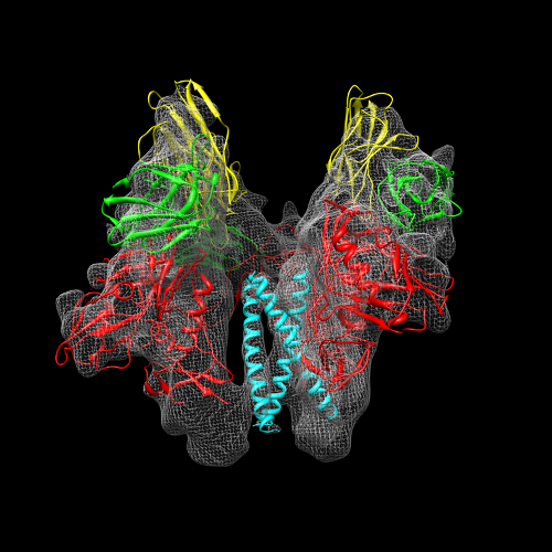 emd_5462.png emd_5462.png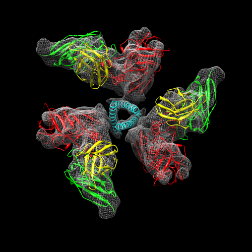 emd_5462_1.png emd_5462_1.png | 225.8 KB 208.2 KB | ||
| Archive directory |  http://ftp.pdbj.org/pub/emdb/structures/EMD-5462ftp://ftp.pdbj.org/pub/emdb/structures/EMD-5462 http://ftp.pdbj.org/pub/emdb/structures/EMD-5462ftp://ftp.pdbj.org/pub/emdb/structures/EMD-5462 | HTTPS FTP |
-Related structure data
| Related structure data |  5455C 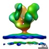 5456C 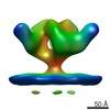 5457C 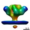 5458C 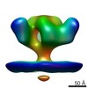 5459C 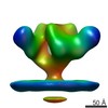 5460C 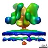 5461C C: citing same article ( |
|---|---|
| Similar structure data |


-Links
| EMDB pages | EMDB (EBI/PDBe) / EMDataResource |
|---|
-Map
| File | Download / File: emd_5462.map.gz / Format: CCP4 / Size: 89 MB / Type: IMAGE STORED AS FLOATING POINT NUMBER (4 BYTES) | ||||||||||||||||||||||||||||||||||||||||||||||||||||||||||||||||||||
|---|---|---|---|---|---|---|---|---|---|---|---|---|---|---|---|---|---|---|---|---|---|---|---|---|---|---|---|---|---|---|---|---|---|---|---|---|---|---|---|---|---|---|---|---|---|---|---|---|---|---|---|---|---|---|---|---|---|---|---|---|---|---|---|---|---|---|---|---|---|
| Annotation | Reconstruction of soluble KNH1144-SOSIP Env trimers in complex with Fab 17b | ||||||||||||||||||||||||||||||||||||||||||||||||||||||||||||||||||||
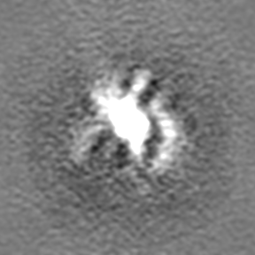
| Projections & slices | Image control
Images are generated by Spider. | ||||||||||||||||||||||||||||||||||||||||||||||||||||||||||||||||||||
| Voxel size | X=Y=Z: 1.08 Å | ||||||||||||||||||||||||||||||||||||||||||||||||||||||||||||||||||||
| Density |
| ||||||||||||||||||||||||||||||||||||||||||||||||||||||||||||||||||||
| Symmetry | Space group: 1 | ||||||||||||||||||||||||||||||||||||||||||||||||||||||||||||||||||||
| Details | EMDB XML:
CCP4 map header:
| ||||||||||||||||||||||||||||||||||||||||||||||||||||||||||||||||||||
 Z (Sec.)
 Y (Row.)
 X (Col.)


-Supplemental data
- Sample components
-Entire : Molecular structure of KNH1144 SOSIP gp140 with 17b Fab.
| Entire | Name: Molecular structure of KNH1144 SOSIP gp140 with 17b Fab. |
|---|---|
| Components |
|
-Supramolecule #1000: Molecular structure of KNH1144 SOSIP gp140 with 17b Fab.
| Supramolecule | Name: Molecular structure of KNH1144 SOSIP gp140 with 17b Fab. type: sample / ID: 1000 / Oligomeric state: trimer / Number unique components: 2 |
|---|---|
| Molecular weight | Theoretical: 670 KDa |
-Macromolecule #1: Envelope glycoprotein
| Macromolecule | Name: Envelope glycoprotein / type: protein_or_peptide / ID: 1 / Name.synonym: Env Details: Membrane proximal external region of KNH1144 (Genbank accession no. AY736812) with the following residue substitutions A662E, S668N, and S676T Number of copies: 3 / Oligomeric state: trimer / Recombinant expression: Yes |
|---|---|
| Source (natural) | Organism: Human immunodeficiency virus 1 / Strain: HIV-1 isolate 00KE_KNH1144 / synonym: HIV-1 |
| Molecular weight | Experimental: 420 KDa |
| Recombinant expression | Organism:  Homo sapiens (human) / Recombinant plasmid: SOSIP-PPI4 and furin-pcDNA3.1 Homo sapiens (human) / Recombinant plasmid: SOSIP-PPI4 and furin-pcDNA3.1 |
-Macromolecule #2: Fab portion of monoclonal antibody 17b
| Macromolecule | Name: Fab portion of monoclonal antibody 17b / type: protein_or_peptide / ID: 2 / Name.synonym: 17b Fab / Details: Fab fragment / Recombinant expression: No / Database: NCBI |
|---|---|
| Source (natural) | Organism: unidentified (others) |
| Molecular weight | Theoretical: 50 KDa |
-Experimental details
-Structure determination
| Method | cryo EM |
|---|---|
 Processing Processing | subtomogram averaging |
| Aggregation state | particle |
-Sample preparation
| Concentration | 0.42 mg/mL |
|---|---|
| Buffer | pH: 7.5 / Details: TNE Buffer (10 mM Tris, 150 mM NaCl, 1 mM EDTA) |
| Grid | Details: Protochips C-flat R 2/2, plasma cleaned |
| Vitrification | Cryogen name: ETHANE / Chamber humidity: 100 % / Instrument: FEI VITROBOT MARK III Method: blot for 6 seconds, blot offset of -2, plunge into an ethane slurry cooled by liquid nitrogen |
- Electron microscopy
| Microscope | FEI TITAN KRIOS |
|---|---|
| Date | Aug 17, 2011 |
| Image recording | Category: CCD / Film or detector model: GATAN ULTRASCAN 4000 (4k x 4k) / Number real images: 3299 / Average electron dose: 20 e/Å2 / Bits/pixel: 16 |
| Electron beam | Acceleration voltage: 80 kV / Electron source:  FIELD EMISSION GUN FIELD EMISSION GUN |
| Electron optics | Calibrated magnification: 138888 / Illumination mode: FLOOD BEAM / Imaging mode: BRIGHT FIELD / Nominal defocus max: 5.0 µm / Nominal defocus min: 1.5 µm / Nominal magnification: 75000 |
| Sample stage | Specimen holder model: FEI TITAN KRIOS AUTOGRID HOLDER |
| Experimental equipment |  Model: Titan Krios / Image courtesy: FEI Company |
-Image processing
| Final reconstruction | Resolution.type: BY AUTHOR / Resolution: 8.8 Å / Resolution method: FSC 0.143 CUT-OFF / Software - Name: EMAN2 / Number subtomograms used: 39001 |
|---|
-Atomic model buiding 1
| Initial model | PDB ID: Chain - #0 - Chain ID: G / Chain - #1 - Chain ID: H / Chain - #2 - Chain ID: L |
|---|---|
| Software | Name: Chimera |
| Details | Protocol: Rigid body. Automated fitting procedures |
| Refinement | Space: REAL / Protocol: RIGID BODY FIT |