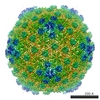
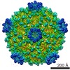
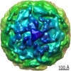
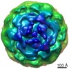
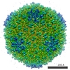
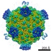
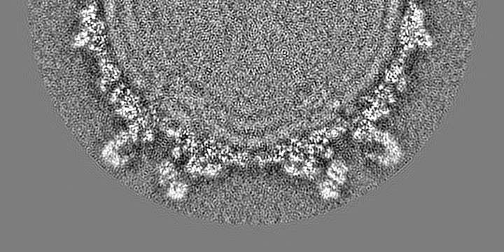
Journal: Proc Natl Acad Sci U S A / Year: 2012 Title: Cryo-EM structure of a transcribing cypovirus. Authors: Chongwen Yang / Gang Ji / Hongrong Liu / Kai Zhang / Guangqiao Liu / Fei Sun / Ping Zhu / Lingpeng Cheng / Abstract: Double-stranded RNA viruses in the family Reoviridae are capable of transcribing and capping nascent mRNA within an icosahedral viral capsid that remains intact throughout repeated transcription ...Double-stranded RNA viruses in the family Reoviridae are capable of transcribing and capping nascent mRNA within an icosahedral viral capsid that remains intact throughout repeated transcription cycles. However, how the highly coordinated mRNA transcription and capping process is facilitated by viral capsid proteins is still unknown. Cypovirus provides a good model system for studying the mRNA transcription and capping mechanism of viruses in the family Reoviridae. Here, we report a full backbone model of a transcribing cypovirus built from a near-atomic-resolution density map by cryoelectron microscopy. Compared with the structure of a nontranscribing cypovirus, the major capsid proteins of transcribing cypovirus undergo a series of conformational changes, giving rise to structural changes in the capsid shell: (i) an enlarged capsid chamber, which provides genomic RNA with more flexibility to move within the densely packed capsid, and (ii) a widened peripentonal channel in the capsid shell, which we confirmed to be a pathway for nascent mRNA. A rod-like structure attributable to a partially resolved nascent mRNA was observed in this channel. In addition, conformational change in the turret protein results in a relatively open turret at each fivefold axis. A GMP moiety, which is transferred to 5'-diphosphorylated mRNA during the mRNA capping reaction, was identified in the pocket-like guanylyltransferase domain of the turret protein.
History
Deposition
Dec 25, 2011
-
Header (metadata) release
Jan 11, 2012
-
Map release
Mar 26, 2012
-
Update
Oct 24, 2012
-
Current status
Oct 24, 2012
Processing site: RCSB / Status: Released
-
Structure visualization
Movie
Surface view with section colored by density value
Specimen holder: Titan Krios / Specimen holder model: OTHER
Experimental equipment
Model: Titan Krios / Image courtesy: FEI Company
-
Image processing
CTF correction
Details: each image
Final reconstruction
Algorithm: OTHER / Resolution.type: BY AUTHOR / Resolution: 4.1 Å / Resolution method: OTHER / Software - Name: IMIRS and ISAF / Number images used: 8000
+
About Yorodumi
-
News
-
Feb 9, 2022. New format data for meta-information of EMDB entries
New format data for meta-information of EMDB entries
Version 3 of the EMDB header file is now the official format.
The previous official version 1.9 will be removed from the archive.
In the structure databanks used in Yorodumi, some data are registered as the other names, "COVID-19 virus" and "2019-nCoV". Here are the details of the virus and the list of structure data.
Jan 31, 2019. EMDB accession codes are about to change! (news from PDBe EMDB page)
EMDB accession codes are about to change! (news from PDBe EMDB page)
The allocation of 4 digits for EMDB accession codes will soon come to an end. Whilst these codes will remain in use, new EMDB accession codes will include an additional digit and will expand incrementally as the available range of codes is exhausted. The current 4-digit format prefixed with “EMD-” (i.e. EMD-XXXX) will advance to a 5-digit format (i.e. EMD-XXXXX), and so on. It is currently estimated that the 4-digit codes will be depleted around Spring 2019, at which point the 5-digit format will come into force.
The EM Navigator/Yorodumi systems omit the EMD- prefix.
Related info.:Q: What is EMD? / ID/Accession-code notation in Yorodumi/EM Navigator
Yorodumi is a browser for structure data from EMDB, PDB, SASBDB, etc.
This page is also the successor to EM Navigator detail page, and also detail information page/front-end page for Omokage search.
The word "yorodu" (or yorozu) is an old Japanese word meaning "ten thousand". "mi" (miru) is to see.
Related info.:EMDB / PDB / SASBDB / Comparison of 3 databanks / Yorodumi Search / Aug 31, 2016. New EM Navigator & Yorodumi / Yorodumi Papers / Jmol/JSmol / Function and homology information / Changes in new EM Navigator and Yorodumi
 Movie
Movie Controller

 Yorodumi
 Open data
 Basic information
 Map data
 Sample
 Keywords
 Function and homology information
 Bombyx mori cypovirus 1
 Authors
 Citation
 Structure visualization
 Downloads & links
 emd_5376_1.jpg
 http://ftp.pdbj.org/pub/emdb/structures/EMD-5376


 Z (Sec.)
 Y (Row.)
 X (Col.)


 Sample components
 Processing
 Electron microscopy
 FIELD EMISSION GUN
