 Movie
Movie Controller


+ Open data

- Basic information
| Entry |  | |||||||||
|---|---|---|---|---|---|---|---|---|---|---|
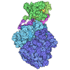
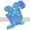
| Title | Map of M. pneumoniae 50S membrane complex in native cells | |||||||||
 Map data Map data | ||||||||||
 Sample Sample |
| |||||||||
 Keywords Keywords | ribosome / TRANSLATION | |||||||||
| Function / homology |  Function and homology information Function and homology informationtransferase activity / large ribosomal subunit / 5S rRNA binding / ribosomal large subunit assembly / large ribosomal subunit rRNA binding / tRNA binding / cytosolic large ribosomal subunit / cytoplasmic translation / response to antibiotic / rRNA binding ...transferase activity / large ribosomal subunit / 5S rRNA binding / ribosomal large subunit assembly / large ribosomal subunit rRNA binding / tRNA binding / cytosolic large ribosomal subunit / cytoplasmic translation / response to antibiotic / rRNA binding / negative regulation of translation / ribonucleoprotein complex / structural constituent of ribosome / ribosome / translation / mRNA binding / plasma membrane / cytoplasm Similarity search - Function | |||||||||
| Biological species |  Mycoplasmoides pneumoniae M129 (bacteria) Mycoplasmoides pneumoniae M129 (bacteria) | |||||||||
| Method | subtomogram averaging / cryo EM / Resolution: 7.7 Å | |||||||||
 Authors Authors | Dobbs JM / Jensen RK / Mahamid J | |||||||||
| Funding support |  United States, United States,  Denmark, 2 items Denmark, 2 items
| |||||||||
 Citation Citation | Journal: Cell / Year: 2026 Title: Single-cell visual proteomics of a minimal bacterium reveals structural coordination of gene expression machineries. Authors: Joseph M Dobbs / Rasmus K Jensen / Julia Mahamid /  Abstract: Translation is a central process in gene expression. Its regulation is complex, depends on factors that include cell state and the subcellular environment, and is subject to modulation via crosstalk ...Translation is a central process in gene expression. Its regulation is complex, depends on factors that include cell state and the subcellular environment, and is subject to modulation via crosstalk to processes such as transcription or translocation. Here, we used cryo-electron tomography of native and antibiotic-perturbed Mycoplasma pneumoniae cells to resolve 140 maps that recapitulate bacterial translation during the initiation, elongation, and recycling phases. We visualized multiple transcription-translation complexes, allowing us to propose a threading-based translation reinitiation mechanism and to provide structural evidence for a long-hypothesized supercomplex that coordinates transcription, translation, and membrane attachment. We resolved abundant membrane-associated large ribosomal subunits and suggest that dissociation from membranes depends on the conditional initiation of new translation, consistent with a potentially conserved mechanism in mammalian cells. This work visualizes the multilayered control of bacterial translation and demonstrates the power of in-cell structural biology to investigate regulatory circuits in gene expression. | |||||||||
| History |
|
- Structure visualization
| Supplemental images |
|---|

- Downloads & links
-EMDB archive
| Map data | emd_53630.map.gz | 90.5 MB | EMDB map data format | |
|---|---|---|---|---|
| Header (meta data) | emd-53630-v30.xmlemd-53630.xml | 16.2 KB 16.2 KB | Display Display | EMDB header |
| FSC (resolution estimation) | emd_53630_fsc.xml | 12.1 KB | Display | FSC data file |
| Images |  emd_53630.png emd_53630.png | 35.5 KB | ||
| Filedesc metadata | emd-53630.cif.gz | 4.5 KB | ||
| Others | emd_53630_half_map_1.map.gzemd_53630_half_map_2.map.gz | 52.9 MB 52.9 MB | ||
| Archive directory |  https://data.pdbj.org/pub/emdb/structures/EMD-53630ftp://data.pdbj.org/pub/emdb/structures/EMD-53630 https://data.pdbj.org/pub/emdb/structures/EMD-53630ftp://data.pdbj.org/pub/emdb/structures/EMD-53630 | HTTPS FTP |
-Related structure data
| Related structure data |  9skdMC  9sk2C  9sk4C  9sk5C  9sk6C  9sk7C  9sk8C  9sk9C  9skaC  9skbC  9skcC  9skeC  9skfC  9sl4C  9sl5C  9sl6C  9sl7C M: atomic model generated by this map C: citing same article ( |
|---|---|
| Similar structure data |


-Links
| EMDB pages | EMDB (EBI/PDBe) / EMDataResource |
|---|---|
| Related items in Molecule of the Month |
-Map
| File | Download / File: emd_53630.map.gz / Format: CCP4 / Size: 103 MB / Type: IMAGE STORED AS FLOATING POINT NUMBER (4 BYTES) | ||||||||||||||||||||||||||||||||||||
|---|---|---|---|---|---|---|---|---|---|---|---|---|---|---|---|---|---|---|---|---|---|---|---|---|---|---|---|---|---|---|---|---|---|---|---|---|---|
| Projections & slices | Image control
Images are generated by Spider. | ||||||||||||||||||||||||||||||||||||
| Voxel size | X=Y=Z: 2 Å | ||||||||||||||||||||||||||||||||||||
| Density |
| ||||||||||||||||||||||||||||||||||||
| Symmetry | Space group: 1 | ||||||||||||||||||||||||||||||||||||
| Details | EMDB XML:
|
 Z (Sec.)
 Y (Row.)
 X (Col.)


-Supplemental data
-Half map: #1
| File | emd_53630_half_map_1.map | ||||||||||||
|---|---|---|---|---|---|---|---|---|---|---|---|---|---|
| Projections & Slices |
| ||||||||||||
| Density Histograms |


-Half map: #2
| File | emd_53630_half_map_2.map | ||||||||||||
|---|---|---|---|---|---|---|---|---|---|---|---|---|---|
| Projections & Slices |
| ||||||||||||
| Density Histograms |


- Sample components
-Entire : Mycoplasma pneumoniae M129-B7
| Entire | Name: Mycoplasma pneumoniae M129-B7 |
|---|---|
| Components |
|
-Supramolecule #1: Mycoplasma pneumoniae M129-B7
| Supramolecule | Name: Mycoplasma pneumoniae M129-B7 / type: cell / ID: 1 / Parent: 0 |
|---|---|
| Source (natural) | Organism: Mycoplasmoides pneumoniae M129 (bacteria) |
-Experimental details
-Structure determination
| Method | cryo EM |
|---|---|
 Processing Processing | subtomogram averaging |
| Aggregation state | cell |
-Sample preparation
| Buffer | pH: 7.4 |
|---|---|
| Grid | Model: Quantifoil R2/1 / Material: GOLD / Mesh: 200 / Support film - Material: CARBON / Support film - topology: HOLEY |
| Vitrification | Cryogen name: ETHANE-PROPANE |
- Electron microscopy #1
| Microscopy ID | 1 |
|---|---|
| Microscope | TFS KRIOS |
| Specialist optics | Phase plate: VOLTA PHASE PLATE |
| Image recording | Image recording ID: 1 / Film or detector model: GATAN K3 BIOQUANTUM (6k x 4k) / Detector mode: COUNTING / Average electron dose: 3.2 e/Å2 |
| Electron beam | Acceleration voltage: 300 kV / Electron source:  FIELD EMISSION GUN FIELD EMISSION GUN |
| Electron optics | Calibrated magnification: 53000 / Illumination mode: FLOOD BEAM / Imaging mode: BRIGHT FIELD / Nominal defocus max: 5.0 µm / Nominal defocus min: 2.5 µm |
| Experimental equipment |  Model: Titan Krios / Image courtesy: FEI Company |
-Electron microscopy #1~
| Microscopy ID | 1 |
|---|---|
| Microscope | TFS KRIOS |
| Specialist optics | Phase plate: VOLTA PHASE PLATE |
| Image recording | Image recording ID: 2 / Film or detector model: GATAN K2 SUMMIT (4k x 4k) / Detector mode: COUNTING / Average electron dose: 3.2 e/Å2 |
| Electron beam | Acceleration voltage: 300 kV / Electron source: FIELD EMISSION GUN |
| Electron optics | Calibrated magnification: 53000 / Illumination mode: FLOOD BEAM / Imaging mode: BRIGHT FIELD / Nominal defocus max: 5.0 µm / Nominal defocus min: 2.5 µm |
| Experimental equipment | Model: Titan Krios / Image courtesy: FEI Company |

