Movie
Movie Controller

[English] 日本語
 Yorodumi
- EMDB-53590: Structural characterisation of chromatin remodelling intermediate... -
+ Open data

- Basic information
| Entry |  | |||||||||
|---|---|---|---|---|---|---|---|---|---|---|
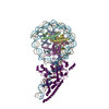
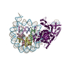
| Title | Structural characterisation of chromatin remodelling intermediates supports linker DNA dependent product inhibition as a mechanism for nucleosome spacing. | |||||||||
 Map data Map data | ||||||||||
 Sample Sample |
| |||||||||
 Keywords Keywords | Nucleosome / Remodelling enzyme / GENE REGULATION | |||||||||
| Function / homology |  Function and homology information Function and homology informationstructural constituent of chromatin / nucleosome / nucleosome assembly / heterochromatin formation / protein heterodimerization activity / DNA binding / nucleoplasm / nucleus Similarity search - Function | |||||||||
| Biological species |  | |||||||||
| Method | single particle reconstruction / cryo EM / Resolution: 4.2 Å | |||||||||
 Authors Authors | Sundaramoorthy R / Hughes A / Owen-hughes TA | |||||||||
| Funding support |  United Kingdom, 1 items United Kingdom, 1 items
| |||||||||
 Citation Citation | Journal: Elife / Year: 2025 Title: Structural characterisation of chromatin remodelling intermediates supports linker DNA-dependent product inhibition as a mechanism for nucleosome spacing. Authors: Amanda L Hughes / Ramasubramanian Sundaramoorthy / Tom Owen-Hughes / Abstract: Previously, we showed that Chd1 chromatin remodelling enzyme associates with nucleosomes oriented towards the longer linker (Sundaramoorthy et al., 2018) (1). Here, we report a series of structures ...Previously, we showed that Chd1 chromatin remodelling enzyme associates with nucleosomes oriented towards the longer linker (Sundaramoorthy et al., 2018) (1). Here, we report a series of structures of Chd1 bound to nucleosomes during ongoing ATP-dependent repositioning. Combining these with biochemical experiments and existing literature, we propose a model in which Chd1 first associates oriented to sample putative entry DNA. In an ATP-dependent reaction, the enzyme then redistributes to the opposite side of the nucleosome, where it subsequently adopts a conformation productive for DNA translocation. Once this active complex extends the nascent exit linker to approximately 15 bp, it is sensed by the Chd1 DNA binding domain, resulting in conversion to a product-inhibited state. These observations provide a mechanistic basis for the action of a molecular ruler element in nucleosome spacing. | |||||||||
| History |
|
- Structure visualization
| Supplemental images |
|---|

- Downloads & links
-EMDB archive
| Map data | emd_53590.map.gz | 595.4 MB | EMDB map data format | |
|---|---|---|---|---|
| Header (meta data) | emd-53590-v30.xmlemd-53590.xml | 30.1 KB 30.1 KB | Display Display | EMDB header |
| FSC (resolution estimation) | emd_53590_fsc.xml | 18.7 KB | Display | FSC data file |
| Images |  emd_53590.png emd_53590.png | 147.7 KB | ||
| Masks | emd_53590_msk_1.map | 669.9 MB | Mask map | |
| Filedesc metadata | emd-53590.cif.gz | 7.3 KB | ||
| Others | emd_53590_additional_1.map.gzemd_53590_half_map_1.map.gzemd_53590_half_map_2.map.gz | 323.7 MB 621 MB 620.8 MB | ||
| Archive directory |  http://ftp.pdbj.org/pub/emdb/structures/EMD-53590ftp://ftp.pdbj.org/pub/emdb/structures/EMD-53590 http://ftp.pdbj.org/pub/emdb/structures/EMD-53590ftp://ftp.pdbj.org/pub/emdb/structures/EMD-53590 | HTTPS FTP |
-Related structure data
| Related structure data |  9r5kMC  9r5sC  9r5wC M: atomic model generated by this map C: citing same article ( |
|---|---|
| Similar structure data |


-Links
| EMDB pages | EMDB (EBI/PDBe) / EMDataResource |
|---|---|
| Related items in Molecule of the Month |


-Map
| File | Download / File: emd_53590.map.gz / Format: CCP4 / Size: 669.9 MB / Type: IMAGE STORED AS FLOATING POINT NUMBER (4 BYTES) | ||||||||||||||||||||||||||||||||||||
|---|---|---|---|---|---|---|---|---|---|---|---|---|---|---|---|---|---|---|---|---|---|---|---|---|---|---|---|---|---|---|---|---|---|---|---|---|---|
| Projections & slices | Image control
Images are generated by Spider. | ||||||||||||||||||||||||||||||||||||
| Voxel size | X=Y=Z: 0.55 Å | ||||||||||||||||||||||||||||||||||||
| Density |
| ||||||||||||||||||||||||||||||||||||
| Symmetry | Space group: 1 | ||||||||||||||||||||||||||||||||||||
| Details | EMDB XML:
|
 Z (Sec.)
 Y (Row.)
 X (Col.)


-Supplemental data
-Mask #1
| File | emd_53590_msk_1.map | ||||||||||||
|---|---|---|---|---|---|---|---|---|---|---|---|---|---|
| Projections & Slices |
| ||||||||||||
| Density Histograms |


-Additional map: #1
| File | emd_53590_additional_1.map | ||||||||||||
|---|---|---|---|---|---|---|---|---|---|---|---|---|---|
| Projections & Slices |
| ||||||||||||
| Density Histograms |


-Half map: #2
| File | emd_53590_half_map_1.map | ||||||||||||
|---|---|---|---|---|---|---|---|---|---|---|---|---|---|
| Projections & Slices |
| ||||||||||||
| Density Histograms |


-Half map: #1
| File | emd_53590_half_map_2.map | ||||||||||||
|---|---|---|---|---|---|---|---|---|---|---|---|---|---|
| Projections & Slices |
| ||||||||||||
| Density Histograms |


- Sample components
-Entire : Nucleosome-Chd1 complex
| Entire | Name: Nucleosome-Chd1 complex |
|---|---|
| Components |
|
-Supramolecule #1: Nucleosome-Chd1 complex
| Supramolecule | Name: Nucleosome-Chd1 complex / type: complex / ID: 1 / Parent: 0 / Macromolecule list: all / Details: Chd1 remodeller bound to Nucleosome |
|---|---|
| Source (natural) | Organism: |
| Molecular weight | Theoretical: 400 KDa |
-Macromolecule #1: DNA (162-MER)
| Macromolecule | Name: DNA (162-MER) / type: dna / ID: 1 / Number of copies: 1 / Classification: DNA |
|---|---|
| Source (natural) | Organism: synthetic construct (others) |
| Molecular weight | Theoretical: 49.864762 KDa |
| Sequence | String: (DC)(DC)(DT)(DA)(DT)(DA)(DC)(DG)(DC)(DG) (DG)(DG)(DC)(DG)(DC)(DA)(DC)(DT)(DG)(DC) (DA)(DG)(DA)(DA)(DG)(DC)(DT)(DT)(DG) (DG)(DT)(DC)(DC)(DC)(DG)(DG)(DG)(DG)(DC) (DC) (DG)(DC)(DT)(DC)(DA)(DA) ...String: (DC)(DC)(DT)(DA)(DT)(DA)(DC)(DG)(DC)(DG) (DG)(DG)(DC)(DG)(DC)(DA)(DC)(DT)(DG)(DC) (DA)(DG)(DA)(DA)(DG)(DC)(DT)(DT)(DG) (DG)(DT)(DC)(DC)(DC)(DG)(DG)(DG)(DG)(DC) (DC) (DG)(DC)(DT)(DC)(DA)(DA)(DT)(DT) (DG)(DG)(DT)(DC)(DG)(DT)(DA)(DG)(DG)(DA) (DA)(DG) (DC)(DT)(DC)(DT)(DA)(DG)(DA) (DT)(DC)(DC)(DG)(DC)(DT)(DT)(DA)(DA)(DA) (DC)(DG)(DA) (DA)(DC)(DG)(DT)(DA)(DC) (DG)(DC)(DG)(DC)(DT)(DG)(DT)(DC)(DC)(DC) (DC)(DC)(DG)(DC) (DG)(DT)(DT)(DT)(DT) (DA)(DA)(DC)(DC)(DG)(DC)(DC)(DA)(DA)(DG) (DG)(DG)(DG)(DA)(DT) (DT)(DA)(DC)(DT) (DC)(DC)(DC)(DT)(DA)(DG)(DT)(DC)(DT)(DC) (DC)(DA)(DG)(DG)(DC)(DA) (DC)(DG)(DT) (DG)(DT)(DC)(DA)(DG)(DA)(DT)(DA)(DT)(DA) (DT)(DA)(DC)(DA)(DT)(DC)(DG) (DA)(DT) |
-Macromolecule #2: DNA (162-MER)
| Macromolecule | Name: DNA (162-MER) / type: dna / ID: 2 / Number of copies: 1 / Classification: DNA |
|---|---|
| Source (natural) | Organism: synthetic construct (others) |
| Molecular weight | Theoretical: 50.153941 KDa |
| Sequence | String: (DA)(DT)(DC)(DG)(DA)(DT)(DG)(DT)(DA)(DT) (DA)(DT)(DA)(DT)(DC)(DT)(DG)(DA)(DC)(DA) (DC)(DG)(DT)(DG)(DC)(DC)(DT)(DG)(DG) (DA)(DG)(DA)(DC)(DT)(DA)(DG)(DG)(DG)(DA) (DG) (DT)(DA)(DA)(DT)(DC)(DC) ...String: (DA)(DT)(DC)(DG)(DA)(DT)(DG)(DT)(DA)(DT) (DA)(DT)(DA)(DT)(DC)(DT)(DG)(DA)(DC)(DA) (DC)(DG)(DT)(DG)(DC)(DC)(DT)(DG)(DG) (DA)(DG)(DA)(DC)(DT)(DA)(DG)(DG)(DG)(DA) (DG) (DT)(DA)(DA)(DT)(DC)(DC)(DC)(DC) (DT)(DT)(DG)(DG)(DC)(DG)(DG)(DT)(DT)(DA) (DA)(DA) (DA)(DC)(DG)(DC)(DG)(DG)(DG) (DG)(DG)(DA)(DC)(DA)(DG)(DC)(DG)(DC)(DG) (DT)(DA)(DC) (DG)(DT)(DT)(DC)(DG)(DT) (DT)(DT)(DA)(DA)(DG)(DC)(DG)(DG)(DA)(DT) (DC)(DT)(DA)(DG) (DA)(DG)(DC)(DT)(DT) (DC)(DC)(DT)(DA)(DC)(DG)(DA)(DC)(DC)(DA) (DA)(DT)(DT)(DG)(DA) (DG)(DC)(DG)(DG) (DC)(DC)(DC)(DC)(DG)(DG)(DG)(DA)(DC)(DC) (DA)(DA)(DG)(DC)(DT)(DT) (DC)(DT)(DG) (DC)(DA)(DG)(DT)(DG)(DC)(DG)(DC)(DC)(DC) (DG)(DC)(DG)(DT)(DA)(DT)(DA) (DG)(DG) |
-Macromolecule #3: Histone H3.2
| Macromolecule | Name: Histone H3.2 / type: protein_or_peptide / ID: 3 / Number of copies: 2 / Enantiomer: LEVO |
|---|---|
| Source (natural) | Organism: |
| Molecular weight | Theoretical: 15.421101 KDa |
| Recombinant expression | Organism:  |
| Sequence | String: MARTKQTARK STGGKAPRKQ LATKAARKSA PATGGVKKPH RYRPGTVALR EIRRYQKSTE LLIRKLPFQR LVREIAQDFK TDLRFQSSA VMALQEASEA YLVGLFEDTN LCAIHAKRVT IMPKDIQLAR RIRGERA UniProtKB: Histone H3.2 |
-Macromolecule #4: Histone H4
| Macromolecule | Name: Histone H4 / type: protein_or_peptide / ID: 4 / Number of copies: 2 / Enantiomer: LEVO |
|---|---|
| Source (natural) | Organism: |
| Molecular weight | Theoretical: 11.394426 KDa |
| Recombinant expression | Organism: |
| Sequence | String: MSGRGKGGKG LGKGGAKRHR KVLRDNIQGI TKPAIRRLAR RGGVKRISGL IYEETRGVLK VFLENVIRDA VTYTEHAKRK TVTAMDVVY ALKRQGRTLY GFGG UniProtKB: Histone H4 |
-Macromolecule #5: Histone H2A type 1
| Macromolecule | Name: Histone H2A type 1 / type: protein_or_peptide / ID: 5 / Number of copies: 2 / Enantiomer: LEVO |
|---|---|
| Source (natural) | Organism: |
| Molecular weight | Theoretical: 13.993295 KDa |
| Recombinant expression | Organism: |
| Sequence | String: MSGRGKQGGK TRAKAKTRSS RAGLQFPVGR VHRLLRKGNY AERVGAGAPV YLAAVLEYLT AEILELAGNA ARDNKKTRII PRHLQLAVR NDEELNKLLG GVTIAQGGVL PNIQSVLLPK KTESAKSAKS K UniProtKB: Histone H2A type 1 |
-Macromolecule #6: Histone H2B 1.1
| Macromolecule | Name: Histone H2B 1.1 / type: protein_or_peptide / ID: 6 / Number of copies: 2 / Enantiomer: LEVO |
|---|---|
| Source (natural) | Organism: |
| Molecular weight | Theoretical: 13.965265 KDa |
| Recombinant expression | Organism: |
| Sequence | String: MPEPAKSAPA PKKGSKKAVT KTQKKDGKKR RKSRKESYAI YVYKVLKQVH PDTGISSKAM SIMNSFVNDV FERIAGEASR LAHYNKRST ITSREIQTAV RLLLPGELAK HAVSEGTKAV TKYTSAK UniProtKB: Histone H2B 1.1 |
-Experimental details
-Structure determination
| Method | cryo EM |
|---|---|
 Processing Processing | single particle reconstruction |
| Aggregation state | particle |
-Sample preparation
| Concentration | 1.5 mg/mL | |||||||||
|---|---|---|---|---|---|---|---|---|---|---|
| Buffer | pH: 7.5 Component:
Details: 20mM Hepes, 120mM Nacl | |||||||||
| Grid | Model: Quantifoil R2/1 / Material: COPPER/RHODIUM / Mesh: 400 / Support film - Material: CARBON / Support film - topology: HOLEY / Pretreatment - Type: GLOW DISCHARGE / Pretreatment - Time: 60 sec. / Pretreatment - Atmosphere: AIR / Pretreatment - Pressure: 0.0001 kPa | |||||||||
| Vitrification | Cryogen name: ETHANE / Chamber humidity: 100 % / Chamber temperature: 277 K / Instrument: FEI VITROBOT MARK IV Details: Vitrified carried out in climate chamber with 100% humidity. | |||||||||
| Details | Purified Nucleosome-Chd1 complex. |
- Electron microscopy
| Microscope | TFS KRIOS |
|---|---|
| Temperature | Min: 77.0 K / Max: 77.0 K |
| Specialist optics | Energy filter - Name: GIF Bioquantum / Energy filter - Slit width: 20 eV |
| Image recording | Film or detector model: GATAN K3 BIOQUANTUM (6k x 4k) / Number grids imaged: 1 / Number real images: 2562 / Average exposure time: 10.0 sec. / Average electron dose: 46.0 e/Å2 |
| Electron beam | Acceleration voltage: 300 kV / Electron source:  FIELD EMISSION GUN FIELD EMISSION GUN |
| Electron optics | C2 aperture diameter: 50.0 µm / Calibrated defocus max: 3.2 µm / Calibrated defocus min: 1.8 µm / Calibrated magnification: 81000 / Illumination mode: FLOOD BEAM / Imaging mode: BRIGHT FIELD / Cs: 2.7 mm / Nominal defocus max: 3.2 µm / Nominal defocus min: 1.8 µm / Nominal magnification: 81000 |
| Sample stage | Specimen holder model: FEI TITAN KRIOS AUTOGRID HOLDER / Cooling holder cryogen: NITROGEN |
| Experimental equipment |  Model: Titan Krios / Image courtesy: FEI Company |
+Image processing


-Atomic model buiding 1
| Initial model |
| ||||||||
|---|---|---|---|---|---|---|---|---|---|
| Refinement | Space: REAL / Protocol: FLEXIBLE FIT / Overall B value: 180 / Target criteria: cross-correlation | ||||||||
| Output model | PDB-9r5k: |