[English] 日本語
 Yorodumi
- EMDB-5341: Chemically controlled assembly of 1, 2 and 3-dimensional protein ... -
+ Open data
- Basic information
| Entry | Database: EMDB / ID: EMD-5341 | |||||||||
|---|---|---|---|---|---|---|---|---|---|---|
| Title | Chemically controlled assembly of 1, 2 and 3-dimensional protein arrays | |||||||||
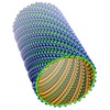
 Map data Map data | This is a surface rendered side-view of an engineered RIDC3 nanotube | |||||||||
 Sample Sample |
| |||||||||
 Keywords Keywords | nanotube / self assembly / protein array / ridc3 / metal coordination | |||||||||
| Function / homology | Cytochrome b562 / Cytochrome b562 / Cytochrome c/b562 / electron transport chain / periplasmic space / electron transfer activity / iron ion binding / heme binding / Soluble cytochrome b562 Function and homology information Function and homology information | |||||||||
| Biological species |  | |||||||||
| Method | helical reconstruction / cryo EM / Resolution: 10.0 Å | |||||||||
 Authors Authors | Brodin JD / Ambroggio XI / Tang C / Parent KN / Baker TS / Tezcan FA | |||||||||
 Citation Citation |  Journal: Nat Chem / Year: 2012 Journal: Nat Chem / Year: 2012Title: Metal-directed, chemically tunable assembly of one-, two- and three-dimensional crystalline protein arrays. Authors: Jeffrey D Brodin / X I Ambroggio / Chunyan Tang / Kristin N Parent / Timothy S Baker / F Akif Tezcan /  Abstract: Proteins represent the most sophisticated building blocks available to an organism and to the laboratory chemist. Yet, in contrast to nearly all other types of molecular building blocks, the designed ...Proteins represent the most sophisticated building blocks available to an organism and to the laboratory chemist. Yet, in contrast to nearly all other types of molecular building blocks, the designed self-assembly of proteins has largely been inaccessible because of the chemical and structural heterogeneity of protein surfaces. To circumvent the challenge of programming extensive non-covalent interactions to control protein self-assembly, we have previously exploited the directionality and strength of metal coordination interactions to guide the formation of closed, homoligomeric protein assemblies. Here, we extend this strategy to the generation of periodic protein arrays. We show that a monomeric protein with properly oriented coordination motifs on its surface can arrange, on metal binding, into one-dimensional nanotubes and two- or three-dimensional crystalline arrays with dimensions that collectively span nearly the entire nano- and micrometre scale. The assembly of these arrays is tuned predictably by external stimuli, such as metal concentration and pH. | |||||||||
| History |
|
- Structure visualization
| Movie |
 Movie viewer Movie viewer |
|---|---|
| Structure viewer | EM map:  SurfView SurfView Molmil Molmil Jmol/JSmol Jmol/JSmol |
| Supplemental images |
- Downloads & links
-EMDB archive
| Map data |  emd_5341.map.gz emd_5341.map.gz | 57.9 MB |  EMDB map data format EMDB map data format | |
|---|---|---|---|---|
| Header (meta data) |  emd-5341-v30.xml emd-5341-v30.xml emd-5341.xml emd-5341.xml | 10 KB 10 KB | Display Display |  EMDB header EMDB header |
| Images |  emd_5341_1.png emd_5341_1.png | 477.2 KB | ||
| Archive directory |  http://ftp.pdbj.org/pub/emdb/structures/EMD-5341 http://ftp.pdbj.org/pub/emdb/structures/EMD-5341 ftp://ftp.pdbj.org/pub/emdb/structures/EMD-5341 ftp://ftp.pdbj.org/pub/emdb/structures/EMD-5341 | HTTPS FTP |
-Validation report
| Summary document |  emd_5341_validation.pdf.gz emd_5341_validation.pdf.gz | 78.9 KB | Display |  EMDB validaton report EMDB validaton report |
|---|---|---|---|---|
| Full document |  emd_5341_full_validation.pdf.gz emd_5341_full_validation.pdf.gz | 78 KB | Display | |
| Data in XML |  emd_5341_validation.xml.gz emd_5341_validation.xml.gz | 493 B | Display | |
| Arichive directory |  https://ftp.pdbj.org/pub/emdb/validation_reports/EMD-5341 https://ftp.pdbj.org/pub/emdb/validation_reports/EMD-5341 ftp://ftp.pdbj.org/pub/emdb/validation_reports/EMD-5341 ftp://ftp.pdbj.org/pub/emdb/validation_reports/EMD-5341 | HTTPS FTP |
-Related structure data
- Links
Links
| EMDB pages |  EMDB (EBI/PDBe) / EMDB (EBI/PDBe) /  EMDataResource EMDataResource |
|---|---|
| Related items in Molecule of the Month |
- Map
Map
| File |  Download / File: emd_5341.map.gz / Format: CCP4 / Size: 238.4 MB / Type: IMAGE STORED AS FLOATING POINT NUMBER (4 BYTES) Download / File: emd_5341.map.gz / Format: CCP4 / Size: 238.4 MB / Type: IMAGE STORED AS FLOATING POINT NUMBER (4 BYTES) | ||||||||||||||||||||||||||||||||||||||||||||||||||||||||||||||||||||
|---|---|---|---|---|---|---|---|---|---|---|---|---|---|---|---|---|---|---|---|---|---|---|---|---|---|---|---|---|---|---|---|---|---|---|---|---|---|---|---|---|---|---|---|---|---|---|---|---|---|---|---|---|---|---|---|---|---|---|---|---|---|---|---|---|---|---|---|---|---|
| Annotation | This is a surface rendered side-view of an engineered RIDC3 nanotube | ||||||||||||||||||||||||||||||||||||||||||||||||||||||||||||||||||||
| Projections & slices | Image control
Images are generated by Spider. | ||||||||||||||||||||||||||||||||||||||||||||||||||||||||||||||||||||
| Voxel size | X=Y=Z: 2.88 Å | ||||||||||||||||||||||||||||||||||||||||||||||||||||||||||||||||||||
| Density |
| ||||||||||||||||||||||||||||||||||||||||||||||||||||||||||||||||||||
| Symmetry | Space group: 1 | ||||||||||||||||||||||||||||||||||||||||||||||||||||||||||||||||||||
| Details | EMDB XML:
CCP4 map header:
| ||||||||||||||||||||||||||||||||||||||||||||||||||||||||||||||||||||
-Supplemental data
- Sample components
-Entire : an engineered cytochrome cb562, RIDC3
| Entire | Name: an engineered cytochrome cb562, RIDC3 |
|---|---|
| Components |
|
-Supramolecule #1000: an engineered cytochrome cb562, RIDC3
| Supramolecule | Name: an engineered cytochrome cb562, RIDC3 / type: sample / ID: 1000 / Oligomeric state: The asymmetric unit is a tetramer / Number unique components: 1 |
|---|---|
| Molecular weight | Experimental: 124 KDa / Method: MALDI |
-Macromolecule #1: cytochrome cb562 variant
| Macromolecule | Name: cytochrome cb562 variant / type: protein_or_peptide / ID: 1 / Name.synonym: ridc3 / Oligomeric state: dimer / Recombinant expression: Yes |
|---|---|
| Source (natural) | Organism:  |
| Molecular weight | Experimental: 124 KDa / Theoretical: 124 KDa |
| Recombinant expression | Organism:  |
-Experimental details
-Structure determination
| Method | cryo EM |
|---|---|
 Processing Processing | helical reconstruction |
| Aggregation state | filament |
- Sample preparation
| Concentration | 1.2 mg/mL |
|---|---|
| Buffer | pH: 5.5 / Details: 20 mM MES, 10 mM ZnCl2 |
| Grid | Details: home-made lacey carbon with a thin layer of continuous carbon |
| Vitrification | Cryogen name: ETHANE / Chamber humidity: 100 % / Chamber temperature: 89 K / Instrument: HOMEMADE PLUNGER / Details: Vitrification instrument: manual plunge-freezer / Method: Blot for 5 seconds before freezing |
- Electron microscopy
| Microscope | FEI POLARA 300 |
|---|---|
| Temperature | Min: 90 K / Max: 90 K / Average: 90 K |
| Alignment procedure | Legacy - Astigmatism: at working magnification |
| Date | Jun 23, 2011 |
| Image recording | Category: CCD / Film or detector model: GENERIC GATAN (4k x 4k) / Average electron dose: 15 e/Å2 |
| Electron beam | Acceleration voltage: 200 kV / Electron source:  FIELD EMISSION GUN FIELD EMISSION GUN |
| Electron optics | Calibrated magnification: 52027 / Illumination mode: FLOOD BEAM / Imaging mode: BRIGHT FIELD / Cs: 2.3 mm / Nominal defocus max: 3.675 µm / Nominal defocus min: 1.75 µm / Nominal magnification: 39000 |
| Sample stage | Specimen holder: Polara Multi Specimen Holder / Specimen holder model: GATAN LIQUID NITROGEN |
| Experimental equipment |  Model: Tecnai Polara / Image courtesy: FEI Company |
- Image processing
| Final reconstruction | Applied symmetry - Helical parameters - Δz: 25.7 Å Applied symmetry - Helical parameters - Δ&Phi: 5.3 ° Applied symmetry - Helical parameters - Axial symmetry: C9 (9 fold cyclic) Algorithm: OTHER / Resolution.type: BY AUTHOR / Resolution: 10.0 Å / Resolution method: OTHER / Software - Name: IHRSR |
|---|---|
| CTF correction | Details: ROBEM and IHRSR |
 Movie
Movie Controller


 Z (Sec.)
 Y (Row.)
 X (Col.)


