[English] 日本語
Yorodumi
- PDB-3tol: Crystal structure of an engineered cytochrome cb562 that forms 1D... -

+
Open data


ID or keywords:

Loading...

-
Basic information

Entry
Database: PDB / ID: 3tol
TitleCrystal structure of an engineered cytochrome cb562 that forms 1D, Zn-mediated coordination polymers
ComponentsSoluble cytochrome b562
KeywordsMETAL BINDING PROTEIN / four helix bundle / electron transfer / periplasmic space
Function / homology
Function and homology information


electron transfer activity / periplasmic space / iron ion binding / heme binding
Similarity search - Function
Cytochrome c/b562 / Cytochrome b562 / Cytochrome b562 / Cytochrome c/b562 / Four Helix Bundle (Hemerythrin (Met), subunit A) / Up-down Bundle / Mainly Alpha
Similarity search - Domain/homology
PROTOPORPHYRIN IX CONTAINING FE / Soluble cytochrome b562
Similarity search - Component
Biological speciesEscherichia coli (E. coli)
MethodX-RAY DIFFRACTION / SYNCHROTRON / MOLECULAR REPLACEMENT / molecular replacement / Resolution: 2 Å
AuthorsBrodin, J.B. / Tezcan, F.A.
Citation
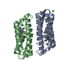
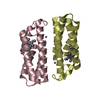
Journal: Nat Chem / Year: 2012
Title: Metal-directed, chemically tunable assembly of one-, two- and three-dimensional crystalline protein arrays.
Authors: Jeffrey D Brodin / X I Ambroggio / Chunyan Tang / Kristin N Parent / Timothy S Baker / F Akif Tezcan /
Abstract: Proteins represent the most sophisticated building blocks available to an organism and to the laboratory chemist. Yet, in contrast to nearly all other types of molecular building blocks, the designed ...Proteins represent the most sophisticated building blocks available to an organism and to the laboratory chemist. Yet, in contrast to nearly all other types of molecular building blocks, the designed self-assembly of proteins has largely been inaccessible because of the chemical and structural heterogeneity of protein surfaces. To circumvent the challenge of programming extensive non-covalent interactions to control protein self-assembly, we have previously exploited the directionality and strength of metal coordination interactions to guide the formation of closed, homoligomeric protein assemblies. Here, we extend this strategy to the generation of periodic protein arrays. We show that a monomeric protein with properly oriented coordination motifs on its surface can arrange, on metal binding, into one-dimensional nanotubes and two- or three-dimensional crystalline arrays with dimensions that collectively span nearly the entire nano- and micrometre scale. The assembly of these arrays is tuned predictably by external stimuli, such as metal concentration and pH.
History
DepositionSep 5, 2011Deposition site: RCSB / Processing site: RCSB
Revision 1.0Jul 4, 2012Provider: repository / Type: Initial release
Revision 1.1Feb 28, 2024Group: Data collection / Database references ...Data collection / Database references / Derived calculations / Refinement description
Category: chem_comp_atom / chem_comp_bond ...chem_comp_atom / chem_comp_bond / database_2 / pdbx_struct_conn_angle / struct_conn / struct_ncs_dom_lim / struct_ref_seq_dif / struct_site
Item: _database_2.pdbx_DOI / _database_2.pdbx_database_accession ..._database_2.pdbx_DOI / _database_2.pdbx_database_accession / _pdbx_struct_conn_angle.ptnr1_auth_asym_id / _pdbx_struct_conn_angle.ptnr1_auth_comp_id / _pdbx_struct_conn_angle.ptnr1_auth_seq_id / _pdbx_struct_conn_angle.ptnr1_label_asym_id / _pdbx_struct_conn_angle.ptnr1_label_atom_id / _pdbx_struct_conn_angle.ptnr1_label_comp_id / _pdbx_struct_conn_angle.ptnr1_label_seq_id / _pdbx_struct_conn_angle.ptnr2_auth_asym_id / _pdbx_struct_conn_angle.ptnr2_auth_comp_id / _pdbx_struct_conn_angle.ptnr2_auth_seq_id / _pdbx_struct_conn_angle.ptnr2_label_asym_id / _pdbx_struct_conn_angle.ptnr2_label_atom_id / _pdbx_struct_conn_angle.ptnr2_label_comp_id / _pdbx_struct_conn_angle.ptnr3_auth_asym_id / _pdbx_struct_conn_angle.ptnr3_auth_comp_id / _pdbx_struct_conn_angle.ptnr3_auth_seq_id / _pdbx_struct_conn_angle.ptnr3_label_asym_id / _pdbx_struct_conn_angle.ptnr3_label_atom_id / _pdbx_struct_conn_angle.ptnr3_label_comp_id / _pdbx_struct_conn_angle.ptnr3_label_seq_id / _pdbx_struct_conn_angle.value / _struct_conn.pdbx_dist_value / _struct_conn.ptnr1_auth_asym_id / _struct_conn.ptnr1_auth_comp_id / _struct_conn.ptnr1_auth_seq_id / _struct_conn.ptnr1_label_asym_id / _struct_conn.ptnr1_label_atom_id / _struct_conn.ptnr1_label_comp_id / _struct_conn.ptnr1_label_seq_id / _struct_conn.ptnr2_auth_asym_id / _struct_conn.ptnr2_auth_comp_id / _struct_conn.ptnr2_auth_seq_id / _struct_conn.ptnr2_label_asym_id / _struct_conn.ptnr2_label_atom_id / _struct_conn.ptnr2_label_comp_id / _struct_conn.ptnr2_label_seq_id / _struct_ncs_dom_lim.beg_auth_comp_id / _struct_ncs_dom_lim.beg_label_asym_id / _struct_ncs_dom_lim.beg_label_comp_id / _struct_ncs_dom_lim.beg_label_seq_id / _struct_ncs_dom_lim.end_auth_comp_id / _struct_ncs_dom_lim.end_label_asym_id / _struct_ncs_dom_lim.end_label_comp_id / _struct_ncs_dom_lim.end_label_seq_id / _struct_ref_seq_dif.details / _struct_site.pdbx_auth_asym_id / _struct_site.pdbx_auth_comp_id / _struct_site.pdbx_auth_seq_id

-
Structure visualization

Structure viewerMolecule:
MolmilJmol/JSmol

Downloads & links

-
Assembly

Deposited unit
A: Soluble cytochrome b562
C: Soluble cytochrome b562
D: Soluble cytochrome b562
B: Soluble cytochrome b562
hetero molecules


Theoretical massNumber of molelcules
Total (without water)49,33614
Polymers46,5284
Non-polymers2,80810
Water6,539363
1
A: Soluble cytochrome b562
B: Soluble cytochrome b562
hetero molecules


Theoretical massNumber of molelcules
Total (without water)24,6687
Polymers23,2642
Non-polymers1,4045
Water362
TypeNameSymmetry operationNumber
identity operation1_555x,y,z1
Buried area3700 Å2
ΔGint-106 kcal/mol
Surface area11030 Å2
MethodPISA
2
C: Soluble cytochrome b562
D: Soluble cytochrome b562
hetero molecules


Theoretical massNumber of molelcules
Total (without water)24,6687
Polymers23,2642
Non-polymers1,4045
Water362
TypeNameSymmetry operationNumber
identity operation1_555x,y,z1
Buried area3780 Å2
ΔGint-105 kcal/mol
Surface area11020 Å2
MethodPISA
Unit cell
Length a, b, c (Å)56.546, 69.506, 126.567
Angle α, β, γ (deg.)90.000, 90.000, 90.000
Int Tables number19
Space group name H-MP212121
Noncrystallographic symmetry (NCS)NCS domain:
IDEns-IDDetails
11A
21C
31D
41B

NCS domain segments:

Component-ID: 1 / Ens-ID: 1 / Beg auth comp-ID: ALA / Beg label comp-ID: ALA / End auth comp-ID: ARG / End label comp-ID: ARG / Refine code: 5 / Auth seq-ID: 1 - 106 / Label seq-ID: 1 - 106

Dom-IDAuth asym-IDLabel asym-ID
1AA
2CB
3DC
4BD

-
Components

#1: Protein
Soluble cytochrome b562 / Cytochrome b-562


Mass: 11632.122 Da / Num. of mol.: 4 / Fragment: Soluble cytochrome b562
Mutation: D73H, K77H, R98C, Y101C, K27E, D28K, T31E, R34L, L38A, Q41L, H59R, D66A, V69M, L76A
Source method: isolated from a genetically manipulated source
Source: (gene. exp.) Escherichia coli (E. coli) / Gene: cybC / Production host: Escherichia coli (E. coli) / References: UniProt: P0ABE7
#2: Chemical
ChemComp-HEM / PROTOPORPHYRIN IX CONTAINING FE / HEME / Heme B


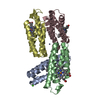
Mass: 616.487 Da / Num. of mol.: 4 / Source method: obtained synthetically / Formula: C34H32FeN4O4
#3: Chemical
ChemComp-ZN / ZINC ION


Mass: 65.409 Da / Num. of mol.: 4 / Source method: obtained synthetically / Formula: Zn
#4: Chemical ChemComp-CA / CALCIUM ION


Mass: 40.078 Da / Num. of mol.: 2 / Source method: obtained synthetically / Formula: Ca
#5: Water ChemComp-HOH / water / Water


Mass: 18.015 Da / Num. of mol.: 363 / Source method: isolated from a natural source / Formula: H2O

-
Experimental details

-
Experiment

ExperimentMethod: X-RAY DIFFRACTION / Number of used crystals: 1

-
Sample preparation

CrystalDensity Matthews: 2.67 Å3/Da / Density % sol: 53.98 %
Crystal growTemperature: 298 K / Method: vapor diffusion, sitting drop / pH: 6.5
Details: 20% PEG 3350, 2.1 mM ZnCl2, 20mM CaCl2 and 100 mM BISTRIS, pH 6.5, VAPOR DIFFUSION, SITTING DROP, temperature 298K

-
Data collection

DiffractionMean temperature: 100 K
Diffraction sourceSource: SYNCHROTRON / Site: SSRL / Beamline: BL7-1 / Wavelength: 0.976 Å
DetectorType: ADSC QUANTUM 315r / Detector: CCD / Date: Aug 4, 2008
RadiationMonochromator: Si(111) / Protocol: SINGLE WAVELENGTH / Monochromatic (M) / Laue (L): M / Scattering type: x-ray
Radiation wavelengthWavelength: 0.976 Å / Relative weight: 1
ReflectionResolution: 2→63.28 Å / Num. all: 34407 / Num. obs: 34063 / % possible obs: 99 % / Observed criterion σ(I): 3
Reflection shellResolution: 2→2.11 Å / Redundancy: 3.6 % / Rmerge(I) obs: 0.349 / Mean I/σ(I) obs: 3.4 / Num. unique all: 4949 / Rsym value: 0.349 / % possible all: 99.8

-
Phasing

PhasingMethod: molecular replacement
Phasing MR
Highest resolutionLowest resolution
Rotation3 Å63.28 Å
Translation3 Å63.28 Å

-
Processing

Software
NameVersionClassificationNB
MOLREPphasing
REFMACrefinement
PDB_EXTRACT3.1data extraction
Blu-Icedata collection
MOSFLMdata reduction
SCALAdata scaling
RefinementMethod to determine structure: MOLECULAR REPLACEMENT / Resolution: 2→63.28 Å / Cor.coef. Fo:Fc: 0.951 / Cor.coef. Fo:Fc free: 0.919 / WRfactor Rfree: 0.2406 / WRfactor Rwork: 0.1871 / Occupancy max: 1 / Occupancy min: 1 / FOM work R set: 0.8459 / SU B: 7.602 / SU ML: 0.116 / SU R Cruickshank DPI: 0.1876 / SU Rfree: 0.1748 / Cross valid method: THROUGHOUT / σ(F): 0 / ESU R Free: 0.175 / Stereochemistry target values: MAXIMUM LIKELIHOOD
Details: HYDROGENS HAVE BEEN ADDED IN THE RIDING POSITIONS U VALUES : WITH TLS ADDED
RfactorNum. reflection% reflectionSelection details
Rfree0.2453 2442 7.2 %RANDOM
Rwork0.19 ---
obs0.1939 34022 98.71 %-
all-34407 --
Solvent computationIon probe radii: 0.8 Å / Shrinkage radii: 0.8 Å / VDW probe radii: 1.4 Å / Solvent model: BABINET MODEL WITH MASK
Displacement parametersBiso max: 65.5 Å2 / Biso mean: 29.6585 Å2 / Biso min: 10.81 Å2
Baniso -1Baniso -2Baniso -3
1--0.07 Å20 Å20 Å2
2---0.14 Å20 Å2
3---0.2 Å2
Refinement stepCycle: LAST / Resolution: 2→63.28 Å
ProteinNucleic acidLigandSolventTotal
Num. atoms3240 0 178 363 3781
Refine LS restraints
Refine-IDTypeDev idealDev ideal targetNumber
X-RAY DIFFRACTIONr_bond_refined_d0.0120.0223480
X-RAY DIFFRACTIONr_angle_refined_deg1.2592.1214740
X-RAY DIFFRACTIONr_dihedral_angle_1_deg4.6655420
X-RAY DIFFRACTIONr_dihedral_angle_2_deg40.8626.75160
X-RAY DIFFRACTIONr_dihedral_angle_3_deg16.45415616
X-RAY DIFFRACTIONr_dihedral_angle_4_deg23.021512
X-RAY DIFFRACTIONr_chiral_restr0.0820.2492
X-RAY DIFFRACTIONr_gen_planes_refined0.0050.0212644
X-RAY DIFFRACTIONr_mcbond_it0.4621.52120
X-RAY DIFFRACTIONr_mcangle_it0.84423352
X-RAY DIFFRACTIONr_scbond_it1.77731360
X-RAY DIFFRACTIONr_scangle_it2.9494.51388
Refine LS restraints NCS

Ens-ID: 1 / Refine-ID: X-RAY DIFFRACTION

Dom-IDAuth asym-IDNumberTypeRms dev position (Å)Weight position
1A424MEDIUM POSITIONAL0.20.5
2C424MEDIUM POSITIONAL0.170.5
3D424MEDIUM POSITIONAL0.140.5
4B424MEDIUM POSITIONAL0.140.5
1A386LOOSE POSITIONAL0.585
2C386LOOSE POSITIONAL0.595
3D386LOOSE POSITIONAL0.585
4B386LOOSE POSITIONAL0.55
1A424MEDIUM THERMAL0.692
2C424MEDIUM THERMAL0.492
3D424MEDIUM THERMAL0.492
4B424MEDIUM THERMAL0.492
1A386LOOSE THERMAL0.8810
2C386LOOSE THERMAL0.7710
3D386LOOSE THERMAL0.7610
4B386LOOSE THERMAL0.8510
LS refinement shellResolution: 2→2.052 Å / Total num. of bins used: 20
RfactorNum. reflection% reflection
Rfree0.291 191 -
Rwork0.22 2288 -
all-2479 -
obs--99.76 %
Refinement TLS params.

Method: refined / Refine-ID: X-RAY DIFFRACTION

IDL112)L122)L132)L222)L232)L332)S11 (Å °)S12 (Å °)S13 (Å °)S21 (Å °)S22 (Å °)S23 (Å °)S31 (Å °)S32 (Å °)S33 (Å °)T112)T122)T132)T222)T232)T332)Origin x (Å)Origin y (Å)Origin z (Å)
13.38890.9487-0.86143.0977-1.19312.5705-0.02710.28410.2764-0.05210.00470.1522-0.1721-0.09410.02240.03020.0062-0.03780.06810.0270.08625.4286-11.1601-21.9009
23.94210.64410.42943.51041.90643.2909-0.08630.261-0.29890.0029-0.02520.01160.16170.02760.11160.0165-0.00940.01010.0531-0.01830.061420.9059-23.2369-22.7318
32.6999-0.34231.10922.5684-1.56794.558-0.08810.1753-0.01480.1630.0566-0.0062-0.53790.36160.03150.2183-0.13340.07140.1028-0.04750.081736.63422.2124-5.1511
43.09770.3891.6693.44231.15154.6322-0.2782-0.31040.01230.11320.26130.0143-0.3977-0.27760.01690.19290.11880.04360.09640.01940.049419.20052.74835.056
Refinement TLS group
IDRefine-IDRefine TLS-IDAuth asym-IDAuth seq-ID
1X-RAY DIFFRACTION1A1 - 106
2X-RAY DIFFRACTION2B1 - 106
3X-RAY DIFFRACTION3C1 - 106
4X-RAY DIFFRACTION4D1 - 106

+
About Yorodumi

-
News

-
Feb 9, 2022. New format data for meta-information of EMDB entries

New format data for meta-information of EMDB entries

  • Version 3 of the EMDB header file is now the official format.
  • The previous official version 1.9 will be removed from the archive.

Related info.:EMDB header

External links:wwPDB to switch to version 3 of the EMDB data model

-
Aug 12, 2020. Covid-19 info

Covid-19 info

URL: https://pdbj.org/emnavi/covid19.php

New page: Covid-19 featured information page in EM Navigator.

Related info.:Covid-19 info / Mar 5, 2020. Novel coronavirus structure data

+
Mar 5, 2020. Novel coronavirus structure data

Novel coronavirus structure data

Related info.:Yorodumi Speices / Aug 12, 2020. Covid-19 info

External links:COVID-19 featured content - PDBj / Molecule of the Month (242):Coronavirus Proteases

+
Jan 31, 2019. EMDB accession codes are about to change! (news from PDBe EMDB page)

EMDB accession codes are about to change! (news from PDBe EMDB page)

  • The allocation of 4 digits for EMDB accession codes will soon come to an end. Whilst these codes will remain in use, new EMDB accession codes will include an additional digit and will expand incrementally as the available range of codes is exhausted. The current 4-digit format prefixed with “EMD-” (i.e. EMD-XXXX) will advance to a 5-digit format (i.e. EMD-XXXXX), and so on. It is currently estimated that the 4-digit codes will be depleted around Spring 2019, at which point the 5-digit format will come into force.
  • The EM Navigator/Yorodumi systems omit the EMD- prefix.

Related info.:Q: What is EMD? / ID/Accession-code notation in Yorodumi/EM Navigator

External links:EMDB Accession Codes are Changing Soon! / Contact to PDBj

+
Jul 12, 2017. Major update of PDB

Major update of PDB

  • wwPDB released updated PDB data conforming to the new PDBx/mmCIF dictionary.
  • This is a major update changing the version number from 4 to 5, and with Remediation, in which all the entries are updated.
  • In this update, many items about electron microscopy experimental information are reorganized (e.g. em_software).
  • Now, EM Navigator and Yorodumi are based on the updated data.

External links:wwPDB Remediation / Enriched Model Files Conforming to OneDep Data Standards Now Available in the PDB FTP Archive

-
Yorodumi

Thousand views of thousand structures

  • Yorodumi is a browser for structure data from EMDB, PDB, SASBDB, etc.
  • This page is also the successor to EM Navigator detail page, and also detail information page/front-end page for Omokage search.
  • The word "yorodu" (or yorozu) is an old Japanese word meaning "ten thousand". "mi" (miru) is to see.

Related info.:EMDB / PDB / SASBDB / Comparison of 3 databanks / Yorodumi Search / Aug 31, 2016. New EM Navigator & Yorodumi / Yorodumi Papers / Jmol/JSmol / Function and homology information / Changes in new EM Navigator and Yorodumi

Read more