 Movie
Movie Controller


+ Open data

- Basic information
| Entry | Database: EMDB / ID: EMD-5334 | |||||||||
|---|---|---|---|---|---|---|---|---|---|---|
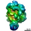
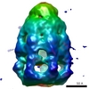
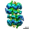
| Title | TRPA1 channel structure at 16A resolution | |||||||||
 Map data Map data | This is a TRPA1 channel map at 16A. | |||||||||
 Sample Sample |
| |||||||||
| Biological species |  | |||||||||
| Method | single particle reconstruction / negative staining / Resolution: 16.0 Å | |||||||||
 Authors Authors | Cvetkov TL / Huynh KW / Cohen MR / Moiseenkova-Bell VY | |||||||||
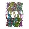
 Citation Citation | Journal: J Biol Chem / Year: 2011 Title: Molecular architecture and subunit organization of TRPA1 ion channel revealed by electron microscopy. Authors: Teresa L Cvetkov / Kevin W Huynh / Matthew R Cohen / Vera Y Moiseenkova-Bell /  Abstract: Transient receptor potential ankyrin 1 (TRPA1) is a non-selective ion channel, which is expressed in nociceptor sensory neurons and transduces chemical, inflammatory, and neuropathic pain signals. ...Transient receptor potential ankyrin 1 (TRPA1) is a non-selective ion channel, which is expressed in nociceptor sensory neurons and transduces chemical, inflammatory, and neuropathic pain signals. Numerous non-reactive compounds and electrophilic compounds, such as endogenous inflammatory mediators and exogenous pungent chemicals, can activate TRPA1. Here we report a 16-Å resolution structure of purified, functional, amphipol-stabilized TRPA1 analyzed by single-particle EM. Molecular models of the N and C termini of the channel were generated using the I-TASSER protein structure prediction server and docked into the EM density to provide insight into the TRPA1 subunit organization. This structural analysis suggests a location for critical N-terminal cysteine residues involved in electrophilic activation at the interface between neighboring subunits. Our results indicate that covalent modifications within this pocket may alter interactions between subunits and promote conformational changes that lead to channel activation. | |||||||||
| History |
|
- Structure visualization
| Movie |
 Movie viewer Movie viewer |
|---|---|
| Structure viewer | EM map: SurfViewMolmilJmol/JSmol |
| Supplemental images |

- Downloads & links
-EMDB archive
| Map data | emd_5334.map.gz | 2.3 MB | EMDB map data format | |
|---|---|---|---|---|
| Header (meta data) | emd-5334-v30.xmlemd-5334.xml | 9.5 KB 9.5 KB | Display Display | EMDB header |
| Images |  emd_5334_1.png emd_5334_1.png | 133.7 KB | ||
| Archive directory |  http://ftp.pdbj.org/pub/emdb/structures/EMD-5334ftp://ftp.pdbj.org/pub/emdb/structures/EMD-5334 http://ftp.pdbj.org/pub/emdb/structures/EMD-5334ftp://ftp.pdbj.org/pub/emdb/structures/EMD-5334 | HTTPS FTP |
-Related structure data
| Similar structure data |
|---|

-Links
| EMDB pages | EMDB (EBI/PDBe) / EMDataResource |
|---|
-Map
| File | Download / File: emd_5334.map.gz / Format: CCP4 / Size: 15.3 MB / Type: IMAGE STORED AS FLOATING POINT NUMBER (4 BYTES) | ||||||||||||||||||||||||||||||||||||||||||||||||||||||||||||||||||||
|---|---|---|---|---|---|---|---|---|---|---|---|---|---|---|---|---|---|---|---|---|---|---|---|---|---|---|---|---|---|---|---|---|---|---|---|---|---|---|---|---|---|---|---|---|---|---|---|---|---|---|---|---|---|---|---|---|---|---|---|---|---|---|---|---|---|---|---|---|---|
| Annotation | This is a TRPA1 channel map at 16A. | ||||||||||||||||||||||||||||||||||||||||||||||||||||||||||||||||||||
| Projections & slices | Image control
Images are generated by Spider. | ||||||||||||||||||||||||||||||||||||||||||||||||||||||||||||||||||||
| Voxel size | X=Y=Z: 3.4 Å | ||||||||||||||||||||||||||||||||||||||||||||||||||||||||||||||||||||
| Density |
| ||||||||||||||||||||||||||||||||||||||||||||||||||||||||||||||||||||
| Symmetry | Space group: 1 | ||||||||||||||||||||||||||||||||||||||||||||||||||||||||||||||||||||
| Details | EMDB XML:
CCP4 map header:
| ||||||||||||||||||||||||||||||||||||||||||||||||||||||||||||||||||||
 Z (Sec.)
 Y (Row.)
 X (Col.)


-Supplemental data
- Sample components
-Entire : TRPA1 channel
| Entire | Name: TRPA1 channel |
|---|---|
| Components |
|
-Supramolecule #1000: TRPA1 channel
| Supramolecule | Name: TRPA1 channel / type: sample / ID: 1000 / Details: The sample was monodisperse. / Oligomeric state: Tetramer / Number unique components: 1 |
|---|---|
| Molecular weight | Experimental: 536 KDa / Theoretical: 525 KDa / Method: Gel filtration |
-Macromolecule #1: transient receptor potential cation channel subfamily A member 1
| Macromolecule | Name: transient receptor potential cation channel subfamily A member 1 type: protein_or_peptide / ID: 1 / Name.synonym: TRPA1 / Number of copies: 1 / Oligomeric state: Tetramer / Recombinant expression: Yes |
|---|---|
| Source (natural) | Organism: |
| Molecular weight | Experimental: 536 KDa / Theoretical: 525 KDa |
| Recombinant expression | Organism:  |
-Experimental details
-Structure determination
| Method | negative staining |
|---|---|
 Processing Processing | single particle reconstruction |
| Aggregation state | particle |
-Sample preparation
| Concentration | 0.1 mg/mL |
|---|---|
| Buffer | pH: 8 Details: 20 mM HEPES, pH 8.0, 150 mM NaCl, 10% glycerol, 1.0 mM DTT |
| Staining | Type: NEGATIVE Details: TRPA1 was adsorbed on carbon-film coated copper grids, washed with 3 droplets of pure water and subsequently stained with 1% uranyl acetate. |
| Grid | Details: 400 mesh copper grids with carbon |
| Vitrification | Cryogen name: NONE / Instrument: OTHER |
- Electron microscopy
| Microscope | FEI TECNAI F20 |
|---|---|
| Alignment procedure | Legacy - Astigmatism: objective lens astigmatism was corrected at 200,000 times magnification |
| Date | May 12, 2011 |
| Image recording | Category: CCD / Film or detector model: GATAN ULTRASCAN 4000 (4k x 4k) / Average electron dose: 20 e/Å2 |
| Tilt angle min | 0 |
| Tilt angle max | 0 |
| Electron beam | Acceleration voltage: 200 kV / Electron source:  FIELD EMISSION GUN FIELD EMISSION GUN |
| Electron optics | Illumination mode: FLOOD BEAM / Imaging mode: BRIGHT FIELD / Cs: 2.0 mm / Nominal defocus max: 1.8 µm / Nominal defocus min: 0.5 µm / Nominal magnification: 55000 |
| Sample stage | Specimen holder: Eucentric / Specimen holder model: OTHER |
| Experimental equipment |  Model: Tecnai F20 / Image courtesy: FEI Company |
-Image processing
| CTF correction | Details: each image |
|---|---|
| Final reconstruction | Resolution.type: BY AUTHOR / Resolution: 16.0 Å / Resolution method: FSC 0.5 CUT-OFF / Software - Name: EMAN2 / Number images used: 8505 |
| Final two d classification | Number classes: 218 |
