 Movie
Movie Controller


+ Open data

- Basic information
| Entry | Database: EMDB / ID: EMD-5317 | |||||||||
|---|---|---|---|---|---|---|---|---|---|---|
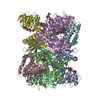
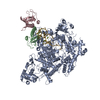
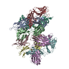
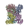
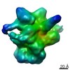
| Title | FcRY dimer | |||||||||
 Map data Map data | This is the cryoEM map of FcRY dimer. | |||||||||
 Sample Sample |
| |||||||||
 Keywords Keywords | avian Fc receptor dimer | |||||||||
| Biological species |  | |||||||||
| Method | single particle reconstruction / cryo EM / negative staining / Resolution: 28.0 Å | |||||||||
 Authors Authors | He Y / Bjorkman PJ | |||||||||
 Citation Citation | Journal: Proc Natl Acad Sci U S A / Year: 2011 Title: Structure of FcRY, an avian immunoglobulin receptor related to mammalian mannose receptors, and its complex with IgY. Authors: Yongning He / Pamela J Bjorkman /  Abstract: Fc receptors transport maternal antibodies across epithelial cell barriers to passively immunize newborns. FcRY, the functional counterpart of mammalian FcRn (a major histocompatibility complex ...Fc receptors transport maternal antibodies across epithelial cell barriers to passively immunize newborns. FcRY, the functional counterpart of mammalian FcRn (a major histocompatibility complex homolog), transfers IgY across the avian yolk sac, and represents a new class of Fc receptor related to the mammalian mannose receptor family. FcRY and FcRn bind immunoglobulins at pH ≤6.5, but not pH ≥7, allowing receptor-ligand association inside intracellular vesicles and release at the pH of blood. We obtained structures of monomeric and dimeric FcRY and an FcRY-IgY complex and explored FcRY's pH-dependent binding mechanism using electron cryomicroscopy (cryoEM) and small-angle X-ray scattering. The cryoEM structure of FcRY at pH 6 revealed a compact double-ring "head," in which the N-terminal cysteine-rich and fibronectin II domains were folded back to contact C-type lectin-like domains 1-6, and a "tail" comprising C-type lectin-like domains 7-8. Conformational changes at pH 8 created a more elongated structure that cannot bind IgY. CryoEM reconstruction of FcRY dimers at pH 6 and small-angle X-ray scattering analysis at both pH values confirmed both structures. The cryoEM structure of the FcRY-IgY revealed symmetric binding of two FcRY heads to the dimeric FcY, each head contacting the C(H)4 domain of one FcY chain. FcRY shares structural properties with mannose receptor family members, including a head and tail domain organization, multimerization that may regulate ligand binding, and pH-dependent conformational changes. Our results facilitate understanding of immune recognition by the structurally related mannose receptor family and comparison of diverse methods of Ig transport across evolution. | |||||||||
| History |
|
- Structure visualization
| Movie |
 Movie viewer Movie viewer |
|---|---|
| Structure viewer | EM map: SurfViewMolmilJmol/JSmol |
| Supplemental images |

- Downloads & links
-EMDB archive
| Map data | emd_5317.map.gz | 383.3 KB | EMDB map data format | |
|---|---|---|---|---|
| Header (meta data) | emd-5317-v30.xmlemd-5317.xml | 8.5 KB 8.5 KB | Display Display | EMDB header |
| Images |  emd_5317_1.png emd_5317_1.png | 67.4 KB | ||
| Archive directory |  http://ftp.pdbj.org/pub/emdb/structures/EMD-5317ftp://ftp.pdbj.org/pub/emdb/structures/EMD-5317 http://ftp.pdbj.org/pub/emdb/structures/EMD-5317ftp://ftp.pdbj.org/pub/emdb/structures/EMD-5317 | HTTPS FTP |
-Related structure data


-Links
| EMDB pages | EMDB (EBI/PDBe) / EMDataResource |
|---|
-Map
| File | Download / File: emd_5317.map.gz / Format: CCP4 / Size: 670.9 KB / Type: IMAGE STORED AS FLOATING POINT NUMBER (4 BYTES) | ||||||||||||||||||||||||||||||||||||||||||||||||||||||||||||||||||||
|---|---|---|---|---|---|---|---|---|---|---|---|---|---|---|---|---|---|---|---|---|---|---|---|---|---|---|---|---|---|---|---|---|---|---|---|---|---|---|---|---|---|---|---|---|---|---|---|---|---|---|---|---|---|---|---|---|---|---|---|---|---|---|---|---|---|---|---|---|---|
| Annotation | This is the cryoEM map of FcRY dimer. | ||||||||||||||||||||||||||||||||||||||||||||||||||||||||||||||||||||
| Projections & slices | Image control
Images are generated by Spider. | ||||||||||||||||||||||||||||||||||||||||||||||||||||||||||||||||||||
| Voxel size | X=Y=Z: 4.24 Å | ||||||||||||||||||||||||||||||||||||||||||||||||||||||||||||||||||||
| Density |
| ||||||||||||||||||||||||||||||||||||||||||||||||||||||||||||||||||||
| Symmetry | Space group: 1 | ||||||||||||||||||||||||||||||||||||||||||||||||||||||||||||||||||||
| Details | EMDB XML:
CCP4 map header:
| ||||||||||||||||||||||||||||||||||||||||||||||||||||||||||||||||||||
 Z (Sec.)
 Y (Row.)
 X (Col.)


-Supplemental data
- Sample components
-Entire : FcRY
| Entire | Name: FcRY |
|---|---|
| Components |
|
-Supramolecule #1000: FcRY
| Supramolecule | Name: FcRY / type: sample / ID: 1000 / Oligomeric state: dimer / Number unique components: 1 |
|---|---|
| Molecular weight | Experimental: 360 KDa |
-Macromolecule #1: FcRY
| Macromolecule | Name: FcRY / type: protein_or_peptide / ID: 1 / Name.synonym: FcRY / Number of copies: 2 / Oligomeric state: dimer / Recombinant expression: Yes |
|---|---|
| Source (natural) | Organism: |
| Molecular weight | Experimental: 180 KDa |
| Recombinant expression | Organism: insect cell (unknown) / Recombinant plasmid: pVL1392 |
-Experimental details
-Structure determination
| Method | negative staining, cryo EM |
|---|---|
 Processing Processing | single particle reconstruction |
| Aggregation state | particle |
-Sample preparation
| Concentration | 2 mg/mL |
|---|---|
| Buffer | pH: 6 / Details: 50 mM Tris, 150 mM NaCl |
| Staining | Type: NEGATIVE / Details: cryo |
| Vitrification | Cryogen name: NITROGEN / Chamber humidity: 100 % / Chamber temperature: 77 K / Instrument: OTHER / Details: Vitrification instrument: FEI vitrobot |
- Electron microscopy
| Microscope | FEI POLARA 300 |
|---|---|
| Temperature | Average: 77 K |
| Specialist optics | Energy filter - Name: Gatan |
| Image recording | Category: CCD / Film or detector model: GATAN ULTRASCAN 1000 (2k x 2k) / Digitization - Sampling interval: 4.24 µm / Average electron dose: 20 e/Å2 |
| Electron beam | Acceleration voltage: 300 kV / Electron source:  FIELD EMISSION GUN FIELD EMISSION GUN |
| Electron optics | Illumination mode: SPOT SCAN / Imaging mode: BRIGHT FIELD / Nominal magnification: 50000 |
| Sample stage | Specimen holder: eucentric / Specimen holder model: OTHER |
| Experimental equipment |  Model: Tecnai Polara / Image courtesy: FEI Company |
-Image processing
| Final reconstruction | Resolution.type: BY AUTHOR / Resolution: 28.0 Å / Resolution method: FSC 0.5 CUT-OFF / Software - Name: EMAN, Frealign / Number images used: 1883 |
|---|
