 Movie
Movie Controller

[English] 日本語
 Yorodumi
- EMDB-52722: Human gamma-tubulin ring complex in centrosomes of HCT116 and HeL... -
+ Open data

- Basic information
| Entry | 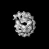 | |||||||||||||||
|---|---|---|---|---|---|---|---|---|---|---|---|---|---|---|---|---|
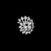
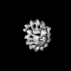
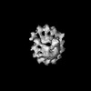
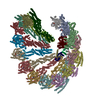
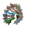
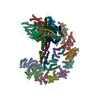
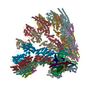
| Title | Human gamma-tubulin ring complex in centrosomes of HCT116 and HeLa cells | |||||||||||||||
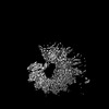
 Map data Map data | Subtomogram average of human gamma-TuRC in centrosomes of HeLa and HCT116 cells | |||||||||||||||
 Sample Sample |
| |||||||||||||||
 Keywords Keywords | centrosome / cytoskeleton / microtubule / microtubule nucleation / complex / template / cap / gamma-tubulin / gamma-tubulin ring complex / cell cycle / nedd1 / neural precursor cell-expressed developmentally down-regulated 1 / CDK5RAP2 / cyclin-dependent kinase 5 regulatory subunit associated protein 2 | |||||||||||||||
| Biological species |  Homo sapiens (human) Homo sapiens (human) | |||||||||||||||
| Method | subtomogram averaging / cryo EM / Resolution: 30.6 Å | |||||||||||||||
 Authors Authors | Hofer FW / Pfeffer S | |||||||||||||||
| Funding support |  Germany, 4 items Germany, 4 items
| |||||||||||||||
 Citation Citation | Journal: Nat Methods / Year: 2023 Title: ELI trifocal microscope: a precise system to prepare target cryo-lamellae for in situ cryo-ET study. Authors: Shuoguo Li / Ziyan Wang / Xing Jia / Tongxin Niu / Jianguo Zhang / Guoliang Yin / Xiaoyun Zhang / Yun Zhu / Gang Ji / Fei Sun /  Abstract: Cryo-electron tomography (cryo-ET) has become a powerful approach to study the high-resolution structure of cellular macromolecular machines in situ. However, the current correlative cryo- ...Cryo-electron tomography (cryo-ET) has become a powerful approach to study the high-resolution structure of cellular macromolecular machines in situ. However, the current correlative cryo-fluorescence and electron microscopy lacks sufficient accuracy and efficiency to precisely prepare cryo-lamellae of target locations for subsequent cryo-ET. Here we describe a precise cryogenic fabrication system, ELI-TriScope, which sets electron (E), light (L) and ion (I) beams at the same focal point to achieve accurate and efficient preparation of a target cryo-lamella. ELI-TriScope uses a commercial dual-beam scanning electron microscope modified to incorporate a cryo-holder-based transfer system and embed an optical imaging system just underneath the vitrified specimen. Cryo-focused ion beam milling can be accurately navigated by monitoring the real-time fluorescence signal of the target molecule. Using ELI-TriScope, we prepared a batch of cryo-lamellae of HeLa cells targeting the centrosome with a success rate of ~91% and discovered new in situ structural features of the human centrosome by cryo-ET. | |||||||||||||||
| History |
|
- Structure visualization
| Supplemental images |
|---|

- Downloads & links
-EMDB archive
| Map data | emd_52722.map.gz | 6 MB |  EMDB map data format EMDB map data format | |
|---|---|---|---|---|
| Header (meta data) | emd-52722-v30.xmlemd-52722.xml | 16.1 KB 16.1 KB | Display Display | EMDB header |
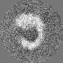
| Images |  emd_52722.png emd_52722.png | 21 KB | ||
| Filedesc metadata | emd-52722.cif.gz | 4.5 KB | ||
| Others | emd_52722_half_map_1.map.gzemd_52722_half_map_2.map.gz | 6 MB 6 MB | ||
| Archive directory |  http://ftp.pdbj.org/pub/emdb/structures/EMD-52722ftp://ftp.pdbj.org/pub/emdb/structures/EMD-52722 http://ftp.pdbj.org/pub/emdb/structures/EMD-52722ftp://ftp.pdbj.org/pub/emdb/structures/EMD-52722 | HTTPS FTP |
-Related structure data
-Links
| EMDB pages | EMDB (EBI/PDBe) / EMDataResource |
|---|
-Map
| File | Download / File: emd_52722.map.gz / Format: CCP4 / Size: 8 MB / Type: IMAGE STORED AS FLOATING POINT NUMBER (4 BYTES) | ||||||||||||||||||||||||||||||||||||
|---|---|---|---|---|---|---|---|---|---|---|---|---|---|---|---|---|---|---|---|---|---|---|---|---|---|---|---|---|---|---|---|---|---|---|---|---|---|
| Annotation | Subtomogram average of human gamma-TuRC in centrosomes of HeLa and HCT116 cells | ||||||||||||||||||||||||||||||||||||
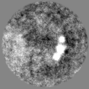
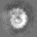
| Projections & slices | Image control
Images are generated by Spider. | ||||||||||||||||||||||||||||||||||||
| Voxel size | X=Y=Z: 4.3015 Å | ||||||||||||||||||||||||||||||||||||
| Density |
| ||||||||||||||||||||||||||||||||||||
| Symmetry | Space group: 1 | ||||||||||||||||||||||||||||||||||||
| Details | EMDB XML:
|
 Z (Sec.)
 Y (Row.)
 X (Col.)


-Supplemental data
-Half map: Half map for subtomogram average of human gamma-TuRC...
| File | emd_52722_half_map_1.map | ||||||||||||
|---|---|---|---|---|---|---|---|---|---|---|---|---|---|
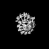
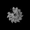
| Annotation | Half map for subtomogram average of human gamma-TuRC in centrosomes of HeLa and HCT116 cells | ||||||||||||
| Projections & Slices |
| ||||||||||||
| Density Histograms |


-Half map: Half map for subtomogram average of human gamma-TuRC...
| File | emd_52722_half_map_2.map | ||||||||||||
|---|---|---|---|---|---|---|---|---|---|---|---|---|---|
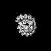
| Annotation | Half map for subtomogram average of human gamma-TuRC in centrosomes of HeLa and HCT116 cells | ||||||||||||
| Projections & Slices |
| ||||||||||||
| Density Histograms |


- Sample components
-Entire : gamma-tubulin ring complex in human centrosomes of HCT116 and HeL...
| Entire | Name: gamma-tubulin ring complex in human centrosomes of HCT116 and HeLa cells |
|---|---|
| Components |
|
-Supramolecule #1: gamma-tubulin ring complex in human centrosomes of HCT116 and HeL...
| Supramolecule | Name: gamma-tubulin ring complex in human centrosomes of HCT116 and HeLa cells type: cell / ID: 1 / Parent: 0 Details: Data from HeLa cells were obtained from EMPIARC-200003 dataset of human centrosomes |
|---|---|
| Source (natural) | Organism: Homo sapiens (human) / Strain: HCT116 & HeLa |
-Experimental details
-Structure determination
| Method | cryo EM |
|---|---|
 Processing Processing | subtomogram averaging |
| Aggregation state | cell |
-Sample preparation
| Buffer | pH: 7 |
|---|---|
| Vitrification | Cryogen name: ETHANE |
- Electron microscopy
| Microscope | TFS KRIOS |
|---|---|
| Image recording | Film or detector model: GATAN K3 BIOQUANTUM (6k x 4k) / Average electron dose: 3.3 e/Å2 |
| Electron beam | Acceleration voltage: 300 kV / Electron source:  FIELD EMISSION GUN FIELD EMISSION GUN |
| Electron optics | Illumination mode: FLOOD BEAM / Imaging mode: BRIGHT FIELD / Nominal defocus max: 6.4 µm / Nominal defocus min: 6.1000000000000005 µm |
| Experimental equipment |  Model: Titan Krios / Image courtesy: FEI Company |
-Image processing
| Final reconstruction | Applied symmetry - Point group: C1 (asymmetric) / Resolution.type: BY AUTHOR / Resolution: 30.6 Å / Resolution method: FSC 0.143 CUT-OFF / Software - Name: RELION (ver. 3.1) / Number subtomograms used: 562 |
|---|---|
| Extraction | Number tomograms: 14 / Number images used: 8260 / Software: (Name: PyTom (ver. 0.971), Warp (ver. 1.09)) |
| Final 3D classification | Number classes: 10 / Software - Name: RELION (ver. 3.1) |
| Final angle assignment | Type: MAXIMUM LIKELIHOOD / Software - Name: RELION (ver. 3.1) |
