ムービー
ムービー コントローラー


+ データを開く

- 基本情報
基本情報
| 登録情報 |  | ||||||||||||
|---|---|---|---|---|---|---|---|---|---|---|---|---|---|
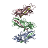
| タイトル | Csu pilus rod type 1 stack | ||||||||||||
 マップデータ マップデータ | Csu pilus type 1 antiparallel stack map, sharpened, pixel spacing adjusted, cropped around the model | ||||||||||||
 試料 試料 |
| ||||||||||||
 キーワード キーワード | pili / fimbriae / Acinetobacter baumannii pili / chaperone-usher pathway / archaic chaperone-usher pili / biofilm / 3D biofilm / adhesion / pathogenesis / pilus antiparallel binding junction / CELL ADHESION | ||||||||||||
| 機能・相同性 | : / Spore coat protein U / Spore Coat Protein U domain / Spore Coat Protein U domain / Spore coat protein U/FanG domain-containing protein 機能・相同性情報 機能・相同性情報 | ||||||||||||
| 生物種 |  Acinetobacter baumannii (バクテリア) Acinetobacter baumannii (バクテリア) | ||||||||||||
| 手法 | 単粒子再構成法 / クライオ電子顕微鏡法 / 解像度: 7.58 Å | ||||||||||||
 データ登録者 データ登録者 | Malmi H / Pakharukova N / Zavialov AV | ||||||||||||
| 資金援助 |  フィンランド, 3件 フィンランド, 3件
| ||||||||||||
 引用 引用 | ジャーナル: To Be Published タイトル: Antiparallel stacking of Csu pili drives Acinetobacter baumannii 3D biofilm assembly 著者: Malmi H / Pakharukova N / Paul B / Tuittila M / Ahmad I / Knight SD / Uhlin BE / Ghosal D / Zavialov AV | ||||||||||||
| 履歴 |
|
- 構造の表示
構造の表示
| 添付画像 |
|---|

- ダウンロードとリンク
-EMDBアーカイブ
| マップデータ | emd_52601.map.gz | 46.7 MB | EMDBマップデータ形式 | |
|---|---|---|---|---|
| ヘッダ (付随情報) | emd-52601-v30.xmlemd-52601.xml | 30.1 KB 30.1 KB | 表示 表示 | EMDBヘッダ |
| FSC (解像度算出) | emd_52601_fsc.xml | 24 KB | 表示 | FSCデータファイル |
| 画像 |  emd_52601.png emd_52601.png | 100.9 KB | ||
| マスクデータ | emd_52601_msk_1.map | 1.4 GB | マスクマップ | |
| Filedesc metadata | emd-52601.cif.gz | 7.9 KB | ||
| その他 | emd_52601_additional_1.map.gzemd_52601_additional_2.map.gzemd_52601_half_map_1.map.gzemd_52601_half_map_2.map.gz | 696.2 MB 716.7 MB 1.3 GB 1.3 GB | ||
| アーカイブディレクトリ |  http://ftp.pdbj.org/pub/emdb/structures/EMD-52601ftp://ftp.pdbj.org/pub/emdb/structures/EMD-52601 http://ftp.pdbj.org/pub/emdb/structures/EMD-52601ftp://ftp.pdbj.org/pub/emdb/structures/EMD-52601 | HTTPS FTP |
-検証レポート
| 文書・要旨 | emd_52601_validation.pdf.gz | 1.1 MB | 表示 | EMDB検証レポート |
|---|---|---|---|---|
| 文書・詳細版 | emd_52601_full_validation.pdf.gz | 1.1 MB | 表示 | |
| XML形式データ | emd_52601_validation.xml.gz | 30.1 KB | 表示 | |
| CIF形式データ | emd_52601_validation.cif.gz | 39.7 KB | 表示 | |
| アーカイブディレクトリ | https://ftp.pdbj.org/pub/emdb/validation_reports/EMD-52601ftp://ftp.pdbj.org/pub/emdb/validation_reports/EMD-52601 | HTTPS FTP |
-関連構造データ


-リンク
| EMDBのページ | EMDB (EBI/PDBe) / EMDataResource |
|---|
-マップ
| ファイル | ダウンロード / ファイル: emd_52601.map.gz / 形式: CCP4 / 大きさ: 54.7 MB / タイプ: IMAGE STORED AS FLOATING POINT NUMBER (4 BYTES) | ||||||||||||||||||||||||||||||||||||
|---|---|---|---|---|---|---|---|---|---|---|---|---|---|---|---|---|---|---|---|---|---|---|---|---|---|---|---|---|---|---|---|---|---|---|---|---|---|
| 注釈 | Csu pilus type 1 antiparallel stack map, sharpened, pixel spacing adjusted, cropped around the model | ||||||||||||||||||||||||||||||||||||
| 投影像・断面図 | 画像のコントロール
画像は Spider により作成 これらの図は立方格子座標系で作成されたものです | ||||||||||||||||||||||||||||||||||||
| ボクセルのサイズ | X=Y=Z: 0.82313 Å | ||||||||||||||||||||||||||||||||||||
| 密度 |
| ||||||||||||||||||||||||||||||||||||
| 対称性 | 空間群: 1 | ||||||||||||||||||||||||||||||||||||
| 詳細 | EMDB XML:
|
 X (Sec.)
 Y (Row.)
 Z (Col.)


-添付データ
-マスク #1
| ファイル | emd_52601_msk_1.map | ||||||||||||
|---|---|---|---|---|---|---|---|---|---|---|---|---|---|
| 投影像・断面図 |
| ||||||||||||
| 密度ヒストグラム |


-追加マップ: Csu pilus type 1 antiparallel stack map, not...
| ファイル | emd_52601_additional_1.map | ||||||||||||
|---|---|---|---|---|---|---|---|---|---|---|---|---|---|
| 注釈 | Csu pilus type 1 antiparallel stack map, not sharpened, pixel spacing adjusted, not cropped around the model | ||||||||||||
| 投影像・断面図 |
| ||||||||||||
| 密度ヒストグラム |


-追加マップ: Csu pilus type 1 antiparallel stack map, sharpened,...
| ファイル | emd_52601_additional_2.map | ||||||||||||
|---|---|---|---|---|---|---|---|---|---|---|---|---|---|
| 注釈 | Csu pilus type 1 antiparallel stack map, sharpened, pixel spacing adjusted, not cropped around the model | ||||||||||||
| 投影像・断面図 |
| ||||||||||||
| 密度ヒストグラム |


-ハーフマップ: Csu pilus type 1 antiparallel stack map, half...
| ファイル | emd_52601_half_map_1.map | ||||||||||||
|---|---|---|---|---|---|---|---|---|---|---|---|---|---|
| 注釈 | Csu pilus type 1 antiparallel stack map, half map A, pixel spacing adjusted | ||||||||||||
| 投影像・断面図 |
| ||||||||||||
| 密度ヒストグラム |


-ハーフマップ: Csu pilus type 1 antiparallel stack map, half...
| ファイル | emd_52601_half_map_2.map | ||||||||||||
|---|---|---|---|---|---|---|---|---|---|---|---|---|---|
| 注釈 | Csu pilus type 1 antiparallel stack map, half map B, pixel spacing adjusted | ||||||||||||
| 投影像・断面図 |
| ||||||||||||
| 密度ヒストグラム |


- 試料の構成要素
-全体 : Csu pilus rod type 1 stack
| 全体 | 名称: Csu pilus rod type 1 stack |
|---|---|
| 要素 |
|
-超分子 #1: Csu pilus rod type 1 stack
| 超分子 | 名称: Csu pilus rod type 1 stack / タイプ: complex / ID: 1 / 親要素: 0 / 含まれる分子: all 詳細: Csu pilus rods are homopolymers of subunit CsuA/B. The sample contains Csu pilus rods self-assembled into type 1 and type 2 stack architectures through antiparallel interactions. The stacks ...詳細: Csu pilus rods are homopolymers of subunit CsuA/B. The sample contains Csu pilus rods self-assembled into type 1 and type 2 stack architectures through antiparallel interactions. The stacks accumulate slowly over time as a gel-like substance at the bottom of a sample tube containing purified Csu pili. |
|---|---|
| 由来(天然) | 生物種: Acinetobacter baumannii (バクテリア) / 株: 19096 / 組織: Planktonic bacteria / Organelle: Outer membrane / 細胞中の位置: Outer membrane |
| 分子量 | 理論値: 16.05974 kDa/nm |
-分子 #1: CsuA/B
| 分子 | 名称: CsuA/B / タイプ: protein_or_peptide / ID: 1 詳細: Csu pilus rods are homopolymers of subunit CsuA/B. Structure depicts three Csu pilus rods forming type 1 stack architecture through antiparallel interactions. コピー数: 57 / 光学異性体: LEVO |
|---|---|
| 由来(天然) | 生物種: Acinetobacter baumannii (バクテリア) / 株: 19096 |
| 分子量 | 理論値: 16.069642 KDa |
| 組換発現 | 生物種: |
| 配列 | 文字列: AVTGQVDVKL NISTGCTVGG SQTEGNMNKF GTLNFGKTSG TWNNVLTAEV ASAATGGNIS VTCDGTDPVD FTVAIDGGER TDRTLKNTA SADVVAYNVY RDAARTNLYV VNQPQQFTTV SGQATAVPIF GAIAPNTGTP KAQGDYKDTL LVTVNF UniProtKB: Spore coat protein U/FanG domain-containing protein |
-実験情報
-構造解析
| 手法 | クライオ電子顕微鏡法 |
|---|---|
 解析 解析 | 単粒子再構成法 |
| 試料の集合状態 | 2D array |
-試料調製
| 緩衝液 | pH: 7.4 構成要素:
詳細: Sample is a fraction taken from an ion exchange column elution gradient, so the NaCl concentration may vary. | |||||||||
|---|---|---|---|---|---|---|---|---|---|---|
| グリッド | モデル: Quantifoil R1.2/1.3 / 材質: COPPER / メッシュ: 300 / 支持フィルム - 材質: CARBON / 支持フィルム - トポロジー: HOLEY / 支持フィルム - Film thickness: 20 / 前処理 - タイプ: GLOW DISCHARGE / 前処理 - 時間: 20 sec. / 前処理 - 雰囲気: AIR / 前処理 - 気圧: 0.014 kPa | |||||||||
| 凍結 | 凍結剤: ETHANE / チャンバー内湿度: 100 % / チャンバー内温度: 277.15 K / 装置: FEI VITROBOT MARK IV | |||||||||
| 詳細 | Csu pilus rods are homopolymers of subunit CsuA/B. The sample contains Csu pilus rods self-assembled into type 1 and type 2 stack architectures through antiparallel interactions. The stacks accumulate slowly over time as a gel-like substance at the bottom of a sample tube containing purified Csu pili. |
- 電子顕微鏡法
| 顕微鏡 | TFS KRIOS |
|---|---|
| 撮影 | フィルム・検出器のモデル: GATAN K3 BIOQUANTUM (6k x 4k) デジタル化 - サイズ - 横: 5760 pixel / デジタル化 - サイズ - 縦: 4092 pixel / 撮影したグリッド数: 1 / 実像数: 7941 / 平均露光時間: 2.79 sec. / 平均電子線量: 59.848 e/Å2 詳細: Grid squares and holes were selected manually as the pilus stacks were visible in the atlas. |
| 電子線 | 加速電圧: 300 kV / 電子線源:  FIELD EMISSION GUN FIELD EMISSION GUN |
| 電子光学系 | C2レンズ絞り径: 50.0 µm / 照射モード: FLOOD BEAM / 撮影モード: BRIGHT FIELD / Cs: 2.7 mm / 最大 デフォーカス(公称値): 1.6 µm / 最小 デフォーカス(公称値): 0.4 µm / 倍率(公称値): 105000 |
| 試料ステージ | 試料ホルダーモデル: FEI TITAN KRIOS AUTOGRID HOLDER ホルダー冷却材: NITROGEN |
| 実験機器 |  モデル: Titan Krios / 画像提供: FEI Company |
+画像解析

-原子モデル構築 1
| 初期モデル | PDB ID: Chain - Residue range: 24-178 / Chain - Source name: PDB / Chain - Initial model type: experimental model 詳細: The initial model consisted of a single pilus rod fragment of the same type. The initial model in turn is based on a crystal structure of CsuA/Bsc with PDB accession code 6FM5. |
|---|---|
| 詳細 | UCSF Chimera was used to first fit the subunits into the single native pilus rod map (PDB Accession ID: 9I37) and average their relative angles, and it was also used to adjust pixel spacing of all maps to match model dimensions. Coot was used to adjust side chain and loop positions. A pair of antiparallel pili with subunit angles adjusted to 3-turns-per-7-subunits symmetry was overlapped with a 2D class to adjust the binding junction angle. Finally, Phenix was used to fit the type 1 stack model into the map. |
| 精密化 | 空間: REAL / プロトコル: RIGID BODY FIT / 温度因子: 668.1 |
| 得られたモデル |  PDB-9i3n: |