Movie
Movie Controller


+ Open data

- Basic information
| Entry |  | ||||||||||||
|---|---|---|---|---|---|---|---|---|---|---|---|---|---|
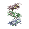
| Title | Csu pilus rod antiparallel pair | ||||||||||||
 Map data Map data | Csu pilus rod pair map, sharpened, pixel spacing adjusted, cropped around the model | ||||||||||||
 Sample Sample |
| ||||||||||||
 Keywords Keywords | pili / fimbriae / Acinetobacter baumannii pili / chaperone-usher pathway / archaic chaperone-usher pili / biofilm / 3D biofilm / adhesion / pathogenesis / pilus antiparallel binding junction / CELL ADHESION | ||||||||||||
| Function / homology | : / Spore coat protein U / Spore Coat Protein U domain / Spore Coat Protein U domain / Spore coat protein U/FanG domain-containing protein Function and homology information Function and homology information | ||||||||||||
| Biological species |  Acinetobacter baumannii (bacteria) Acinetobacter baumannii (bacteria) | ||||||||||||
| Method | single particle reconstruction / cryo EM / Resolution: 8.91 Å | ||||||||||||
 Authors Authors | Malmi H / Pakharukova N / Zavialov AV | ||||||||||||
| Funding support |  Finland, 3 items Finland, 3 items
| ||||||||||||
 Citation Citation | Journal: Nat Commun / Year: 2026 Title: Antiparallel stacking of Csu pili drives Acinetobacter baumannii 3D biofilm assembly. Authors: Henri Malmi / Natalia Pakharukova / Bindusmita Paul / Minna Tuittila / Irfan Ahmad / Stefan David Knight / Bernt Eric Uhlin / Debnath Ghosal / Anton V Zavialov /    Abstract: Many Gram-negative nosocomial pathogens rely on adhesive filaments, known as archaic chaperone-usher pili, to establish stress- and drug-resistant, multi-layered biofilms. Here, we uncover the ...Many Gram-negative nosocomial pathogens rely on adhesive filaments, known as archaic chaperone-usher pili, to establish stress- and drug-resistant, multi-layered biofilms. Here, we uncover the mechanism by which these pili build three-dimensional (3D) biofilm architectures. In situ analyses of Acinetobacter baumannii biofilms using electron microscopy (EM) reveal an extensive network of ultrathin, flat stacks of archaic Csu pili interconnecting bacterial cells in 3D space. Cryo-EM structures of a single native pilus, pilus pairs, and two types of multi-pilus stacks show that the pili pack into antiparallel sheets, with their rods connected laterally by junctions at their zigzag corners. This antiparallel arrangement ensures that contacts form primarily between pili from interacting cells rather than pili from the same cell. With a remarkably short helical repeat, archaic chaperone-usher pili spontaneously establish a high density of junctions that determines the biofilm's 3D architecture. Our findings may help develop new therapies against multidrug-resistant bacterial infections by targeting pilus-pilus interactions. | ||||||||||||
| History |
|
- Structure visualization
| Supplemental images |
|---|

- Downloads & links
-EMDB archive
| Map data | emd_52600.map.gz | 2.2 MB | EMDB map data format | |
|---|---|---|---|---|
| Header (meta data) | emd-52600-v30.xmlemd-52600.xml | 28.9 KB 28.9 KB | Display Display | EMDB header |
| FSC (resolution estimation) | emd_52600_fsc.xml | 9.9 KB | Display | FSC data file |
| Images |  emd_52600.png emd_52600.png | 99.1 KB | ||
| Masks | emd_52600_msk_1.map | 103 MB | Mask map | |
| Filedesc metadata | emd-52600.cif.gz | 7.5 KB | ||
| Others | emd_52600_additional_1.map.gzemd_52600_half_map_1.map.gzemd_52600_half_map_2.map.gz | 96.9 MB 95.5 MB 95.5 MB | ||
| Archive directory |  http://ftp.pdbj.org/pub/emdb/structures/EMD-52600ftp://ftp.pdbj.org/pub/emdb/structures/EMD-52600 http://ftp.pdbj.org/pub/emdb/structures/EMD-52600ftp://ftp.pdbj.org/pub/emdb/structures/EMD-52600 | HTTPS FTP |
-Related structure data
| Related structure data |  9i3mMC  9i37C  9i3nC  9i3oC M: atomic model generated by this map C: citing same article ( |
|---|---|
| Similar structure data |


-Links
| EMDB pages | EMDB (EBI/PDBe) / EMDataResource |
|---|
-Map
| File | Download / File: emd_52600.map.gz / Format: CCP4 / Size: 2.5 MB / Type: IMAGE STORED AS FLOATING POINT NUMBER (4 BYTES) | ||||||||||||||||||||||||||||||||||||
|---|---|---|---|---|---|---|---|---|---|---|---|---|---|---|---|---|---|---|---|---|---|---|---|---|---|---|---|---|---|---|---|---|---|---|---|---|---|
| Annotation | Csu pilus rod pair map, sharpened, pixel spacing adjusted, cropped around the model | ||||||||||||||||||||||||||||||||||||
| Projections & slices | Image control
Images are generated by Spider. generated in cubic-lattice coordinate | ||||||||||||||||||||||||||||||||||||
| Voxel size | X=Y=Z: 1.6626 Å | ||||||||||||||||||||||||||||||||||||
| Density |
| ||||||||||||||||||||||||||||||||||||
| Symmetry | Space group: 1 | ||||||||||||||||||||||||||||||||||||
| Details | EMDB XML:
|
 X (Sec.)
 Y (Row.)
 Z (Col.)


-Supplemental data
-Mask #1
| File | emd_52600_msk_1.map | ||||||||||||
|---|---|---|---|---|---|---|---|---|---|---|---|---|---|
| Projections & Slices |
| ||||||||||||
| Density Histograms |


-Additional map: Csu pilus rod pair map, sharpened, pixel spacing...
| File | emd_52600_additional_1.map | ||||||||||||
|---|---|---|---|---|---|---|---|---|---|---|---|---|---|
| Annotation | Csu pilus rod pair map, sharpened, pixel spacing adjusted, not cropped around the model | ||||||||||||
| Projections & Slices |
| ||||||||||||
| Density Histograms |


-Half map: Csu pilus rod pair map, half map B, pixel spacing adjusted
| File | emd_52600_half_map_1.map | ||||||||||||
|---|---|---|---|---|---|---|---|---|---|---|---|---|---|
| Annotation | Csu pilus rod pair map, half map B, pixel spacing adjusted | ||||||||||||
| Projections & Slices |
| ||||||||||||
| Density Histograms |


-Half map: Csu pilus rod pair map, half map A, pixel spacing adjusted
| File | emd_52600_half_map_2.map | ||||||||||||
|---|---|---|---|---|---|---|---|---|---|---|---|---|---|
| Annotation | Csu pilus rod pair map, half map A, pixel spacing adjusted | ||||||||||||
| Projections & Slices |
| ||||||||||||
| Density Histograms |


- Sample components
-Entire : Csu pilus rod antiparallel pair
| Entire | Name: Csu pilus rod antiparallel pair |
|---|---|
| Components |
|
-Supramolecule #1: Csu pilus rod antiparallel pair
| Supramolecule | Name: Csu pilus rod antiparallel pair / type: complex / ID: 1 / Parent: 0 / Macromolecule list: all Details: Csu pilus rods are homopolymers of subunit CsuA/B. The sample contains Csu pilus rods self-assembled into pairs through antiparallel interactions. |
|---|---|
| Source (natural) | Organism: Acinetobacter baumannii (bacteria) / Strain: 19096 / Tissue: Planktonic bacteria / Organelle: Outer membrane / Location in cell: Outer membrane |
| Molecular weight | Theoretical: 16.05974 kDa/nm |
-Macromolecule #1: CsuA/B
| Macromolecule | Name: CsuA/B / type: protein_or_peptide / ID: 1 / Number of copies: 26 / Enantiomer: LEVO |
|---|---|
| Source (natural) | Organism: Acinetobacter baumannii (bacteria) / Strain: 19096 |
| Molecular weight | Theoretical: 16.069642 KDa |
| Recombinant expression | Organism: |
| Sequence | String: AVTGQVDVKL NISTGCTVGG SQTEGNMNKF GTLNFGKTSG TWNNVLTAEV ASAATGGNIS VTCDGTDPVD FTVAIDGGER TDRTLKNTA SADVVAYNVY RDAARTNLYV VNQPQQFTTV SGQATAVPIF GAIAPNTGTP KAQGDYKDTL LVTVNF UniProtKB: Spore coat protein U/FanG domain-containing protein |
-Experimental details
-Structure determination
| Method | cryo EM |
|---|---|
 Processing Processing | single particle reconstruction |
| Aggregation state | filament |
-Sample preparation
| Buffer | pH: 7.4 Component:
Details: Sample is a fraction taken from an ion exchange column elution gradient, so the NaCl concentration may vary. | |||||||||
|---|---|---|---|---|---|---|---|---|---|---|
| Grid | Model: Quantifoil R1.2/1.3 / Material: COPPER / Mesh: 300 / Support film - Material: CARBON / Support film - topology: HOLEY / Support film - Film thickness: 20 / Pretreatment - Type: GLOW DISCHARGE | |||||||||
| Vitrification | Cryogen name: ETHANE / Chamber humidity: 100 % / Chamber temperature: 277.15 K / Instrument: FEI VITROBOT MARK IV | |||||||||
| Details | Csu pilus rods are homopolymers of subunit CsuA/B. The sample contains Csu pilus rods self-assembled into pairs through antiparallel interactions. The pairs and stacks accumulate over time in a sample tube containing purified Csu pili. |
- Electron microscopy
| Microscope | TFS KRIOS |
|---|---|
| Image recording | Film or detector model: GATAN K2 SUMMIT (4k x 4k) / Detector mode: SUPER-RESOLUTION / Number grids imaged: 1 / Average electron dose: 50.0 e/Å2 |
| Electron beam | Acceleration voltage: 300 kV / Electron source:  FIELD EMISSION GUN FIELD EMISSION GUN |
| Electron optics | Illumination mode: FLOOD BEAM / Imaging mode: BRIGHT FIELD / Nominal defocus max: 2.5 µm / Nominal defocus min: 0.8 µm / Nominal magnification: 105000 |
| Sample stage | Specimen holder model: FEI TITAN KRIOS AUTOGRID HOLDER / Cooling holder cryogen: NITROGEN |
| Experimental equipment |  Model: Titan Krios / Image courtesy: FEI Company |
+Image processing

-Atomic model buiding 1
| Initial model | PDB ID: Chain - Residue range: 24-178 / Chain - Source name: PDB / Chain - Initial model type: experimental model Details: The initial model consisted of a single pilus rod fragment of the same type. The initial model in turn is based on a crystal structure of CsuA/Bsc with PDB accession code 6FM5. |
|---|---|
| Details | UCSF Chimera was used to first fit the subunits into the single native pilus rod map (PDB Accession ID: 9I37) and average their relative angles, and it was also used to adjust pixel spacing of all maps to match model dimensions. Coot was used to adjust side chain and loop positions. A pair of antiparallel pili with subunit angles adjusted to 3-turns-per-7-subunits symmetry was overlapped with a 2D class to adjust the binding junction angle. Finally, Phenix was used to fit the pair model into the map. |
| Refinement | Space: REAL / Protocol: RIGID BODY FIT / Overall B value: 304.76 |
| Output model | PDB-9i3m: |