[English] 日本語
Yorodumi
- EMDB-52436: PSMA in complex with nanobody 7 and 8 -

+
Open data


ID or keywords:

Loading...

-
Basic information

Entry
Database: EMDB / ID: EMD-52436
TitlePSMA in complex with nanobody 7 and 8
Map dataPSMA with nanobodies 7 and 8
Sample
  • Complex: PSMA in complex with nanobody 8
    • Protein or peptide: Glutamate carboxypeptidase 2
    • Protein or peptide: Nanobody 7
  • Protein or peptide: Nanobody 8
  • Ligand: 2-acetamido-2-deoxy-beta-D-glucopyranose
  • Ligand: ZINC ION
  • Ligand: CALCIUM ION
  • Ligand: CHLORIDE ION
Keywordscarboxypeptidase complex nanobody / MEMBRANE PROTEIN
Function / homology
Function and homology information


Ac-Asp-Glu binding / tetrahydrofolyl-poly(glutamate) polymer binding / glutamate carboxypeptidase II / folic acid-containing compound metabolic process / C-terminal protein deglutamylation / Aspartate and asparagine metabolism / dipeptidase activity / carboxypeptidase activity / metallocarboxypeptidase activity / peptidase activity ...Ac-Asp-Glu binding / tetrahydrofolyl-poly(glutamate) polymer binding / glutamate carboxypeptidase II / folic acid-containing compound metabolic process / C-terminal protein deglutamylation / Aspartate and asparagine metabolism / dipeptidase activity / carboxypeptidase activity / metallocarboxypeptidase activity / peptidase activity / cell surface / proteolysis / extracellular exosome / metal ion binding / membrane / plasma membrane / cytoplasm
Similarity search - Function
Transferrin receptor-like, dimerisation domain / Transferrin receptor-like, dimerisation domain superfamily / Glutamate carboxypeptidase 2-like / Transferrin receptor-like dimerisation domain / PA domain superfamily / PA domain / PA domain / Peptidase M28 / Peptidase family M28
Similarity search - Domain/homology
Glutamate carboxypeptidase 2
Similarity search - Component
Biological speciesCamelus (mammal) / Homo sapiens (human) / Camelus dromedarius (Arabian camel)
Methodsingle particle reconstruction / cryo EM / Resolution: 3.0 Å
AuthorsAlon G / Zalk R / Huynh TT / Zalutsky MR / Weizmann Y / Zarivach R / Papo N
Funding support Israel, United States, United Kingdom, 6 items
OrganizationGrant numberCountry
Other private846497 Israel
United States - Israel Binational Science Foundation (BSF)2019303 United States
National Institutes of Health/National Institute of General Medical Sciences (NIH/NIGMS)R01GM144393 United States
Rosetrees TrustOoR2022/100004 United Kingdom
Israel Science Foundation1615/19 Israel
Worldwide Cancer Research20-0238 United Kingdom
Citation
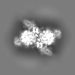
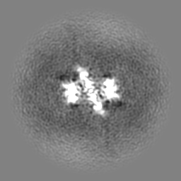
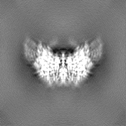
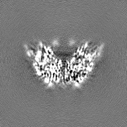
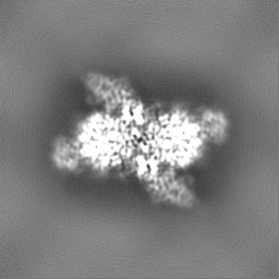
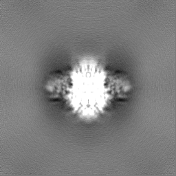
Journal: Int J Biol Macromol / Year: 2025
Title: Structural analysis of nanobody interactions with their prostate-specific membrane antigen binding epitopes.
Authors: Gal Alon-Zchut / Ran Zalk / Truc T Huynh / Michael R Zalutsky / Yossi Weizmann / Raz Zarivach / Niv Papo /
Abstract: Prostate-specific membrane antigen (PSMA), overexpressed in prostate cancer, is a promising target for diagnostics and therapy. However, the monoclonal antibodies in current use for PSMA targeting ...Prostate-specific membrane antigen (PSMA), overexpressed in prostate cancer, is a promising target for diagnostics and therapy. However, the monoclonal antibodies in current use for PSMA targeting and inhibition have suboptimal activities due to their poor tissue and cell penetration and slow normal tissue clearance. Potentially superior alternatives are nanobodies (NBs), the single-chain variable domains of heavy-chain antibodies derived from camelids. The advantages of NBs include small size (~15 kDa), ability to bind hidden epitopes, and rapid clearance. In contrast to most known PSMA inhibitors, which bind to the same catalytic site in PMSA, NBs can bind to different PSMA epitopes, facilitating heterovalent binding strategies that could enhance their therapeutic and diagnostic potential. The objective of this study was to map these binding epitopes and hence to acquire an atomic-resolution understanding of NB-PMSA binding by investigating the structural interactions between PSMA and three NBs (NB7, NB8, and NB37). Using cryo-electron microscopy to generate high-resolution structures of NB-PSMA complexes, we found that NB7 had the highest affinity for PSMA due to a larger interface and to stabilizing interactions, including salt bridges and π-π stacking. Notably, we also found that NB7 and NB8 can bind simultaneously to different PSMA epitopes without interfering with the function of PSMA (which is still not completely known), opening the way for the development of theranostic applications for prostate cancer treatment and imaging. Importantly, NB7 binds specifically to human PSMA but not to murine PSMA, due to key amino acid differences responsible for its species specificity.
History
DepositionDec 29, 2024-
Header (metadata) releaseJul 16, 2025-
Map releaseJul 16, 2025-
UpdateJul 16, 2025-
Current statusJul 16, 2025Processing site: PDBe / Status: Released

-
Structure visualization

Supplemental images

Downloads & links

-
Map

FileDownload / File: emd_52436.map.gz / Format: CCP4 / Size: 64 MB / Type: IMAGE STORED AS FLOATING POINT NUMBER (4 BYTES)
AnnotationPSMA with nanobodies 7 and 8
Projections & slices

Image control

Size
Brightness
Contrast
Others
AxesZ (Sec.)Y (Row.)X (Col.)
0.89 Å/pix.
x 256 pix.
= 227.84 Å
0.89 Å/pix.
x 256 pix.
= 227.84 Å
0.89 Å/pix.
x 256 pix.
= 227.84 Å

Surface

Projections

Slices (1/3)

Slices (1/2)

Slices (2/3)

Images are generated by Spider.

Voxel sizeX=Y=Z: 0.89 Å
Density
Contour LevelBy AUTHOR: 0.06
Minimum - Maximum-0.18248557 - 0.38907444
Average (Standard dev.)0.00041993184 (±0.014985231)
SymmetrySpace group: 1
Details

EMDB XML:

Map geometry
Axis orderXYZ
Origin000
Dimensions256256256
Spacing256256256
CellA=B=C: 227.84 Å
α=β=γ: 90.0 °

-
Supplemental data

-
Half map: PSMA with nanobodies 7 and 8 halfA

Fileemd_52436_half_map_1.map
AnnotationPSMA with nanobodies 7 and 8 halfA
Projections & Slices
AxesZYX

Projections

Slices (1/2)
Density Histograms

-
Half map: PSMA with nanobodies 7 and 8 halfB

Fileemd_52436_half_map_2.map
AnnotationPSMA with nanobodies 7 and 8 halfB
Projections & Slices
AxesZYX

Projections

Slices (1/2)
Density Histograms

-
Sample components

-
Entire : PSMA in complex with nanobody 8

EntireName: PSMA in complex with nanobody 8
Components
  • Complex: PSMA in complex with nanobody 8
    • Protein or peptide: Glutamate carboxypeptidase 2
    • Protein or peptide: Nanobody 7
  • Protein or peptide: Nanobody 8
  • Ligand: 2-acetamido-2-deoxy-beta-D-glucopyranose
  • Ligand: ZINC ION
  • Ligand: CALCIUM ION
  • Ligand: CHLORIDE ION

-
Supramolecule #1: PSMA in complex with nanobody 8

SupramoleculeName: PSMA in complex with nanobody 8 / type: complex / ID: 1 / Parent: 0 / Macromolecule list: #1-#2
Source (natural)Organism: Camelus (mammal)
Molecular weightTheoretical: 230 KDa

-
Macromolecule #1: Glutamate carboxypeptidase 2

MacromoleculeName: Glutamate carboxypeptidase 2 / type: protein_or_peptide / ID: 1 / Number of copies: 2 / Enantiomer: LEVO / EC number: glutamate carboxypeptidase II
Source (natural)Organism: Homo sapiens (human)
Molecular weightTheoretical: 78.341352 KDa
Recombinant expressionOrganism: Escherichia coli (E. coli)
SequenceString: HNMKAFLDEL KAENIKKFLY NFTQIPHLAG TEQNFQLAKQ IQSQWKEFGL DSVELAHYDV LLSYPNKTHP NYISIINEDG NEIFNTSLF EPPPPGYENV SDIVPPFSAF SPQGMPEGDL VYVNYARTED FFKLERDMKI NCSGKIVIAR YGKVFRGNKV K NAQLAGAK ...String:
HNMKAFLDEL KAENIKKFLY NFTQIPHLAG TEQNFQLAKQ IQSQWKEFGL DSVELAHYDV LLSYPNKTHP NYISIINEDG NEIFNTSLF EPPPPGYENV SDIVPPFSAF SPQGMPEGDL VYVNYARTED FFKLERDMKI NCSGKIVIAR YGKVFRGNKV K NAQLAGAK GVILYSDPAD YFAPGVKSYP DGWNLPGGGV QRGNILNLNG AGDPLTPGYP ANEYAYRRGI AEAVGLPSIP VH PIGYYDA QKLLEKMGGS APPDSSWRGS LKVPYNVGPG FTGNFSTQKV KMHIHSTNEV TRIYNVIGTL RGAVEPDRYV ILG GHRDSW VFGGIDPQSG AAVVHEIVRS FGTLKKEGWR PRRTILFASW DAEEFGLLGS TEWAEENSRL LQERGVAYIN ADSS IEGNY TLRVDCTPLM YSLVHNLTKE LKSPDEGFEG KSLYESWTKK SPSPEFSGMP RISKLGSGND FEVFFQRLGI ASGRA RYTK NWETNKFSGY PLYHSVYETY ELVEKFYDPM FKYHLTVAQV RGGMVFELAN SIVLPFDCRD YAVVLRKYAD KIYSIS MKH PQEMKTYSVS FDSLFSAVKN FTEIASKFSE RLQDFDKSNP IVLRMMNDQL MFLERAFIDP LGLPDRPFYR HVIYAPS SH NKYAGESFPG IYDALFDIES KVDPSKAWGE VKRQIYVAAF TVQAAAETLS EVA

UniProtKB: Glutamate carboxypeptidase 2

-
Macromolecule #2: Nanobody 7

MacromoleculeName: Nanobody 7 / type: protein_or_peptide / ID: 2 / Number of copies: 2 / Enantiomer: LEVO
Source (natural)Organism: Camelus dromedarius (Arabian camel)
Molecular weightTheoretical: 13.79518 KDa
Recombinant expressionOrganism: Escherichia coli (E. coli)
SequenceString:
QVQLQESGGG SVQAGGSLRL SCTAPGYTDS NYYMSWFRQA PGKEREWVAG VNTGRGSTSY ADSVKGRFTI SQDNAKNTMF LQMNSLKPE DTAQYYCAVA ACHFCDSLPK TQDEYILWGQ GTQVTVS

-
Macromolecule #3: Nanobody 8

MacromoleculeName: Nanobody 8 / type: protein_or_peptide / ID: 3 / Number of copies: 2 / Enantiomer: LEVO
Source (natural)Organism: Camelus dromedarius (Arabian camel)
Molecular weightTheoretical: 13.299545 KDa
Recombinant expressionOrganism: Escherichia coli (E. coli)
SequenceString:
VQLQESGGGS VQAGGSLRLS CARSGWPYST YSMNWFRQAP GKEREAVAGI SSTMSGIIFA ESKAGQFTIS QDNAKNTVYL QMNNLKPED TAIYYCAARR DYSLSSSSDD FDYWGQGTQV TVS

-
Macromolecule #7: 2-acetamido-2-deoxy-beta-D-glucopyranose

MacromoleculeName: 2-acetamido-2-deoxy-beta-D-glucopyranose / type: ligand / ID: 7 / Number of copies: 8 / Formula: NAG
Molecular weightTheoretical: 221.208 Da
Chemical component information

ChemComp-NAG:
2-acetamido-2-deoxy-beta-D-glucopyranose

-
Macromolecule #8: ZINC ION

MacromoleculeName: ZINC ION / type: ligand / ID: 8 / Number of copies: 4 / Formula: ZN
Molecular weightTheoretical: 65.409 Da

-
Macromolecule #9: CALCIUM ION

MacromoleculeName: CALCIUM ION / type: ligand / ID: 9 / Number of copies: 2 / Formula: CA
Molecular weightTheoretical: 40.078 Da

-
Macromolecule #10: CHLORIDE ION

MacromoleculeName: CHLORIDE ION / type: ligand / ID: 10 / Number of copies: 1 / Formula: CL
Molecular weightTheoretical: 35.453 Da

-
Experimental details

-
Structure determination

Methodcryo EM
Processingsingle particle reconstruction
Aggregation stateparticle

-
Sample preparation

BufferpH: 8
VitrificationCryogen name: ETHANE / Instrument: HOMEMADE PLUNGER / Details: manually blotted for 3 seconds.

-
Electron microscopy

MicroscopeTFS GLACIOS
Image recordingFilm or detector model: TFS FALCON 4i (4k x 4k) / Average electron dose: 30.0 e/Å2
Electron beamAcceleration voltage: 200 kV / Electron source: FIELD EMISSION GUN
Electron opticsIllumination mode: FLOOD BEAM / Imaging mode: BRIGHT FIELD / Nominal defocus max: 2.0 µm / Nominal defocus min: 0.5 µm

+
Image processing

CTF correctionType: NONE
Startup modelType of model: INSILICO MODEL
Final reconstructionAlgorithm: FOURIER SPACE / Resolution.type: BY AUTHOR / Resolution: 3.0 Å / Resolution method: FSC 0.143 CUT-OFF / Number images used: 774655
Initial angle assignmentType: MAXIMUM LIKELIHOOD
Final angle assignmentType: MAXIMUM LIKELIHOOD
FSC plot (resolution estimation)

+
About Yorodumi

-
News

-
Feb 9, 2022. New format data for meta-information of EMDB entries

New format data for meta-information of EMDB entries

  • Version 3 of the EMDB header file is now the official format.
  • The previous official version 1.9 will be removed from the archive.

Related info.:EMDB header

External links:wwPDB to switch to version 3 of the EMDB data model

-
Aug 12, 2020. Covid-19 info

Covid-19 info

URL: https://pdbj.org/emnavi/covid19.php

New page: Covid-19 featured information page in EM Navigator.

Related info.:Covid-19 info / Mar 5, 2020. Novel coronavirus structure data

+
Mar 5, 2020. Novel coronavirus structure data

Novel coronavirus structure data

Related info.:Yorodumi Speices / Aug 12, 2020. Covid-19 info

External links:COVID-19 featured content - PDBj / Molecule of the Month (242):Coronavirus Proteases

+
Jan 31, 2019. EMDB accession codes are about to change! (news from PDBe EMDB page)

EMDB accession codes are about to change! (news from PDBe EMDB page)

  • The allocation of 4 digits for EMDB accession codes will soon come to an end. Whilst these codes will remain in use, new EMDB accession codes will include an additional digit and will expand incrementally as the available range of codes is exhausted. The current 4-digit format prefixed with “EMD-” (i.e. EMD-XXXX) will advance to a 5-digit format (i.e. EMD-XXXXX), and so on. It is currently estimated that the 4-digit codes will be depleted around Spring 2019, at which point the 5-digit format will come into force.
  • The EM Navigator/Yorodumi systems omit the EMD- prefix.

Related info.:Q: What is EMD? / ID/Accession-code notation in Yorodumi/EM Navigator

External links:EMDB Accession Codes are Changing Soon! / Contact to PDBj

+
Jul 12, 2017. Major update of PDB

Major update of PDB

  • wwPDB released updated PDB data conforming to the new PDBx/mmCIF dictionary.
  • This is a major update changing the version number from 4 to 5, and with Remediation, in which all the entries are updated.
  • In this update, many items about electron microscopy experimental information are reorganized (e.g. em_software).
  • Now, EM Navigator and Yorodumi are based on the updated data.

External links:wwPDB Remediation / Enriched Model Files Conforming to OneDep Data Standards Now Available in the PDB FTP Archive

-
Yorodumi

Thousand views of thousand structures

  • Yorodumi is a browser for structure data from EMDB, PDB, SASBDB, etc.
  • This page is also the successor to EM Navigator detail page, and also detail information page/front-end page for Omokage search.
  • The word "yorodu" (or yorozu) is an old Japanese word meaning "ten thousand". "mi" (miru) is to see.

Related info.:EMDB / PDB / SASBDB / Comparison of 3 databanks / Yorodumi Search / Aug 31, 2016. New EM Navigator & Yorodumi / Yorodumi Papers / Jmol/JSmol / Function and homology information / Changes in new EM Navigator and Yorodumi

Read more