 Movie
Movie Controller

[English] 日本語
 Yorodumi
- EMDB-52273: Prostate Specific Membrane Antigen (PSMA) in complex with nanobody7 -
+ Open data

- Basic information
| Entry |  | |||||||||||||||||||||
|---|---|---|---|---|---|---|---|---|---|---|---|---|---|---|---|---|---|---|---|---|---|---|
| Title | Prostate Specific Membrane Antigen (PSMA) in complex with nanobody7 | |||||||||||||||||||||
 Map data Map data | PSMA (prostate specific membrane antigen) in complex with the nanobody NB7 | |||||||||||||||||||||
 Sample Sample |
| |||||||||||||||||||||
 Keywords Keywords | Nanobodies their Prostate Specific Membrane Antigen PSMA prostate cancer / MEMBRANE PROTEIN | |||||||||||||||||||||
| Function / homology |  Function and homology information Function and homology informationintestinal folate absorption / Ac-Asp-Glu binding / tetrahydrofolyl-poly(glutamate) polymer binding / glutamate carboxypeptidase II / folic acid-containing compound metabolic process / Aspartate and asparagine metabolism / dipeptidase activity / : / positive regulation of glutamate receptor signaling pathway / carboxypeptidase activity ...intestinal folate absorption / Ac-Asp-Glu binding / tetrahydrofolyl-poly(glutamate) polymer binding / glutamate carboxypeptidase II / folic acid-containing compound metabolic process / Aspartate and asparagine metabolism / dipeptidase activity / : / positive regulation of glutamate receptor signaling pathway / carboxypeptidase activity / metallocarboxypeptidase activity / peptidase activity / cell surface / proteolysis / extracellular exosome / membrane / metal ion binding / plasma membrane / cytoplasm Similarity search - Function | |||||||||||||||||||||
| Biological species |   Homo sapiens (human) / Homo sapiens (human) / | |||||||||||||||||||||
| Method | single particle reconstruction / cryo EM / Resolution: 2.66 Å | |||||||||||||||||||||
 Authors Authors | Alon G / Papo N / Zarivach R / Zalk R | |||||||||||||||||||||
| Funding support |  United States, United States,  United Kingdom, United Kingdom,  Israel, 6 items Israel, 6 items
| |||||||||||||||||||||
 Citation Citation | Journal: Int J Biol Macromol / Year: 2025 Title: Structural analysis of nanobody interactions with their prostate-specific membrane antigen binding epitopes. Authors: Gal Alon-Zchut / Ran Zalk / Truc T Huynh / Michael R Zalutsky / Yossi Weizmann / Raz Zarivach / Niv Papo / Abstract: Prostate-specific membrane antigen (PSMA), overexpressed in prostate cancer, is a promising target for diagnostics and therapy. However, the monoclonal antibodies in current use for PSMA targeting ...Prostate-specific membrane antigen (PSMA), overexpressed in prostate cancer, is a promising target for diagnostics and therapy. However, the monoclonal antibodies in current use for PSMA targeting and inhibition have suboptimal activities due to their poor tissue and cell penetration and slow normal tissue clearance. Potentially superior alternatives are nanobodies (NBs), the single-chain variable domains of heavy-chain antibodies derived from camelids. The advantages of NBs include small size (~15 kDa), ability to bind hidden epitopes, and rapid clearance. In contrast to most known PSMA inhibitors, which bind to the same catalytic site in PMSA, NBs can bind to different PSMA epitopes, facilitating heterovalent binding strategies that could enhance their therapeutic and diagnostic potential. The objective of this study was to map these binding epitopes and hence to acquire an atomic-resolution understanding of NB-PMSA binding by investigating the structural interactions between PSMA and three NBs (NB7, NB8, and NB37). Using cryo-electron microscopy to generate high-resolution structures of NB-PSMA complexes, we found that NB7 had the highest affinity for PSMA due to a larger interface and to stabilizing interactions, including salt bridges and π-π stacking. Notably, we also found that NB7 and NB8 can bind simultaneously to different PSMA epitopes without interfering with the function of PSMA (which is still not completely known), opening the way for the development of theranostic applications for prostate cancer treatment and imaging. Importantly, NB7 binds specifically to human PSMA but not to murine PSMA, due to key amino acid differences responsible for its species specificity. | |||||||||||||||||||||
| History |
|
- Structure visualization
| Supplemental images |
|---|

- Downloads & links
-EMDB archive
| Map data | emd_52273.map.gz | 31.7 MB | EMDB map data format | |
|---|---|---|---|---|
| Header (meta data) | emd-52273-v30.xmlemd-52273.xml | 20.6 KB 20.6 KB | Display Display | EMDB header |
| FSC (resolution estimation) | emd_52273_fsc.xml | 8.4 KB | Display | FSC data file |
| Images |  emd_52273.png emd_52273.png | 138.2 KB | ||
| Filedesc metadata | emd-52273.cif.gz | 7.3 KB | ||
| Others | emd_52273_half_map_1.map.gzemd_52273_half_map_2.map.gz | 59.3 MB 59.3 MB | ||
| Archive directory |  http://ftp.pdbj.org/pub/emdb/structures/EMD-52273ftp://ftp.pdbj.org/pub/emdb/structures/EMD-52273 http://ftp.pdbj.org/pub/emdb/structures/EMD-52273ftp://ftp.pdbj.org/pub/emdb/structures/EMD-52273 | HTTPS FTP |
-Related structure data
| Related structure data |  9hlwMC  9hviC  9hvkC  9hvlC M: atomic model generated by this map C: citing same article ( |
|---|---|
| Similar structure data |


-Links
| EMDB pages | EMDB (EBI/PDBe) / EMDataResource |
|---|---|
| Related items in Molecule of the Month |

-Map
| File | Download / File: emd_52273.map.gz / Format: CCP4 / Size: 64 MB / Type: IMAGE STORED AS FLOATING POINT NUMBER (4 BYTES) | ||||||||||||||||||||||||||||||||||||
|---|---|---|---|---|---|---|---|---|---|---|---|---|---|---|---|---|---|---|---|---|---|---|---|---|---|---|---|---|---|---|---|---|---|---|---|---|---|
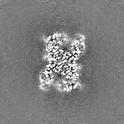
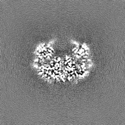
| Annotation | PSMA (prostate specific membrane antigen) in complex with the nanobody NB7 | ||||||||||||||||||||||||||||||||||||
| Projections & slices | Image control
Images are generated by Spider. | ||||||||||||||||||||||||||||||||||||
| Voxel size | X=Y=Z: 0.89 Å | ||||||||||||||||||||||||||||||||||||
| Density |
| ||||||||||||||||||||||||||||||||||||
| Symmetry | Space group: 1 | ||||||||||||||||||||||||||||||||||||
| Details | EMDB XML:
|
 Z (Sec.)
 Y (Row.)
 X (Col.)


-Supplemental data
-Half map: PSMA (prostate specific membrane antigen) in complex with...
| File | emd_52273_half_map_1.map | ||||||||||||
|---|---|---|---|---|---|---|---|---|---|---|---|---|---|
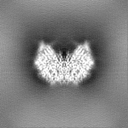
| Annotation | PSMA (prostate specific membrane antigen) in complex with the nanobody NB7 Half map #1 | ||||||||||||
| Projections & Slices |
| ||||||||||||
| Density Histograms |


-Half map: PSMA (prostate specific membrane antigen) in complex with...
| File | emd_52273_half_map_2.map | ||||||||||||
|---|---|---|---|---|---|---|---|---|---|---|---|---|---|
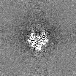
| Annotation | PSMA (prostate specific membrane antigen) in complex with the nanobody NB7 Half map #2 | ||||||||||||
| Projections & Slices |
| ||||||||||||
| Density Histograms |


- Sample components
-Entire : PSMA in complex with nanobody 7
| Entire | Name: PSMA in complex with nanobody 7 |
|---|---|
| Components |
|
-Supramolecule #1: PSMA in complex with nanobody 7
| Supramolecule | Name: PSMA in complex with nanobody 7 / type: complex / ID: 1 / Parent: 0 / Macromolecule list: #1-#2 |
|---|---|
| Source (natural) | Organism: |
| Molecular weight | Theoretical: 230 kDa/nm |
-Macromolecule #1: Glutamate carboxypeptidase 2
| Macromolecule | Name: Glutamate carboxypeptidase 2 / type: protein_or_peptide / ID: 1 / Number of copies: 2 / Enantiomer: LEVO / EC number: glutamate carboxypeptidase II |
|---|---|
| Source (natural) | Organism: Homo sapiens (human) |
| Molecular weight | Theoretical: 84.426445 KDa |
| Recombinant expression | Organism:  |
| Sequence | String: MWNLLHETDS AVATARRPRW LCAGALVLAG GFFLLGFLFG WFIKSSNEAT NITPKHNMKA FLDELKAENI KKFLYNFTQI PHLAGTEQN FQLAKQIQSQ WKEFGLDSVE LAHYDVLLSY PNKTHPNYIS IINEDGNEIF NTSLFEPPPP GYENVSDIVP P FSAFSPQG ...String: MWNLLHETDS AVATARRPRW LCAGALVLAG GFFLLGFLFG WFIKSSNEAT NITPKHNMKA FLDELKAENI KKFLYNFTQI PHLAGTEQN FQLAKQIQSQ WKEFGLDSVE LAHYDVLLSY PNKTHPNYIS IINEDGNEIF NTSLFEPPPP GYENVSDIVP P FSAFSPQG MPEGDLVYVN YARTEDFFKL ERDMKINCSG KIVIARYGKV FRGNKVKNAQ LAGAKGVILY SDPADYFAPG VK SYPDGWN LPGGGVQRGN ILNLNGAGDP LTPGYPANEY AYRRGIAEAV GLPSIPVHPI GYYDAQKLLE KMGGSAPPDS SWR GSLKVP YNVGPGFTGN FSTQKVKMHI HSTNEVTRIY NVIGTLRGAV EPDRYVILGG HRDSWVFGGI DPQSGAAVVH EIVR SFGTL KKEGWRPRRT ILFASWDAEE FGLLGSTEWA EENSRLLQER GVAYINADSS IEGNYTLRVD CTPLMYSLVH NLTKE LKSP DEGFEGKSLY ESWTKKSPSP EFSGMPRISK LGSGNDFEVF FQRLGIASGR ARYTKNWETN KFSGYPLYHS VYETYE LVE KFYDPMFKYH LTVAQVRGGM VFELANSIVL PFDCRDYAVV LRKYADKIYS ISMKHPQEMK TYSVSFDSLF SAVKNFT EI ASKFSERLQD FDKSNPIVLR MMNDQLMFLE RAFIDPLGLP DRPFYRHVIY APSSHNKYAG ESFPGIYDAL FDIESKVD P SKAWGEVKRQ IYVAAFTVQA AAETLSEVA UniProtKB: Glutamate carboxypeptidase 2 |
-Macromolecule #2: Nano body 7
| Macromolecule | Name: Nano body 7 / type: protein_or_peptide / ID: 2 / Number of copies: 2 / Enantiomer: LEVO |
|---|---|
| Source (natural) | Organism: |
| Molecular weight | Theoretical: 16.066593 KDa |
| Recombinant expression | Organism: |
| Sequence | String: QVQLQESGGG SVQAGGSLRL SCTAPGYTDS NYYMSWFRQA PGKEREWVAG VNTGRGSTSY ADSVKGRFTI SQDNAKNTMF LQMNSLKPE DTAIYYCAVA ACHFCDSLPK TQDEYILWGQ GTQVTVSSAA AYPYDVPDYG SHHHHHH |
-Macromolecule #6: 2-acetamido-2-deoxy-beta-D-glucopyranose
| Macromolecule | Name: 2-acetamido-2-deoxy-beta-D-glucopyranose / type: ligand / ID: 6 / Number of copies: 8 / Formula: NAG |
|---|---|
| Molecular weight | Theoretical: 221.208 Da |
| Chemical component information |  ChemComp-NAG: |
-Macromolecule #7: ZINC ION
| Macromolecule | Name: ZINC ION / type: ligand / ID: 7 / Number of copies: 4 / Formula: ZN |
|---|---|
| Molecular weight | Theoretical: 65.409 Da |
-Macromolecule #8: CALCIUM ION
| Macromolecule | Name: CALCIUM ION / type: ligand / ID: 8 / Number of copies: 2 / Formula: CA |
|---|---|
| Molecular weight | Theoretical: 40.078 Da |
-Macromolecule #9: CHLORIDE ION
| Macromolecule | Name: CHLORIDE ION / type: ligand / ID: 9 / Number of copies: 1 / Formula: CL |
|---|---|
| Molecular weight | Theoretical: 35.453 Da |
-Experimental details
-Structure determination
| Method | cryo EM |
|---|---|
 Processing Processing | single particle reconstruction |
| Aggregation state | particle |
-Sample preparation
| Concentration | 1.0 mg/mL |
|---|---|
| Buffer | pH: 8 |
| Grid | Model: Quantifoil R1.2/1.3 / Material: COPPER / Mesh: 200 / Support film - Material: CARBON / Support film - topology: HOLEY / Pretreatment - Type: GLOW DISCHARGE / Pretreatment - Time: 60 sec. |
| Vitrification | Cryogen name: ETHANE Details: Homemade plunger, manually blotted for 3 seconds.. |
- Electron microscopy
| Microscope | TFS GLACIOS |
|---|---|
| Image recording | Film or detector model: TFS FALCON 4i (4k x 4k) / Average electron dose: 30.0 e/Å2 |
| Electron beam | Acceleration voltage: 200 kV / Electron source:  FIELD EMISSION GUN FIELD EMISSION GUN |
| Electron optics | Illumination mode: FLOOD BEAM / Imaging mode: BRIGHT FIELD / Nominal defocus max: 2.0 µm / Nominal defocus min: 0.5 µm |
| Sample stage | Cooling holder cryogen: NITROGEN |


