 Movie
Movie Controller


+ Open data

- Basic information
| Entry | Database: EMDB / ID: EMD-5239 | |||||||||
|---|---|---|---|---|---|---|---|---|---|---|
| Title | human Low Density Lipoprotein | |||||||||
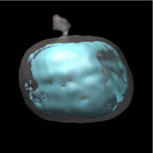
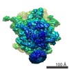
 Map data Map data | Iso-surface rendering of the overall structure of the human LDL particle | |||||||||
 Sample Sample |
| |||||||||
 Keywords Keywords | LDL / cholesterol ester / apoB / atherosclerosis | |||||||||
| Biological species | unidentified (others) | |||||||||
| Method | single particle reconstruction / cryo EM / Resolution: 26.0 Å | |||||||||
 Authors Authors | Liu Y / Atkinson D | |||||||||
 Citation Citation | Journal: J Mol Biol / Year: 2011 Title: Enhancing the contrast of ApoB to locate the surface components in the 3D density map of human LDL. Authors: Yuhang Liu / David Atkinson /  Abstract: A 26 Å resolution map of the structure of human low-density lipoprotein (LDL) was obtained from electron cryomicroscopy and single-particle image reconstruction. The structure showed a discoidal- ...A 26 Å resolution map of the structure of human low-density lipoprotein (LDL) was obtained from electron cryomicroscopy and single-particle image reconstruction. The structure showed a discoidal-shaped LDL particle with high-density regions mainly distributed at the edge of the particle and low-density regions at the flat surface that covers the core region. To determine the chemical components that correspond to these density regions and to delineate the distribution of protein and phospholipid located at the particle surface at the resolution of the map, we used Mono-Sulfo-NHS-Undecagold labeling to increase preferentially the contrast of the apolipoprotein B component on the LDL particle. In the three-dimensional map from the image reconstruction of the undecagold-labeled LDL particles, the high-density region from the undecagold label was distributed mainly at the edge of the particle, and lower density regions were found at the flat surfaces that cover the neutral lipid core. This suggests that apolipoprotein B mainly encircles LDL at the edge of the particle and the phospholipid monolayers are located at the flat surfaces, which are parallel to the cholesterol ester layers in the core and may interact with the core lipid layers through the acyl chains. | |||||||||
| History |
|
- Structure visualization
| Movie |
 Movie viewer Movie viewer |
|---|---|
| Structure viewer | EM map: SurfViewMolmilJmol/JSmol |
| Supplemental images |
- Downloads & links
-EMDB archive
| Map data | emd_5239.map.gz | 684.5 KB | EMDB map data format | |
|---|---|---|---|---|
| Header (meta data) | emd-5239-v30.xmlemd-5239.xml | 9 KB 9 KB | Display Display | EMDB header |
| Images |  emd_5239_1.jpg emd_5239_1.jpg | 18.5 KB | ||
| Archive directory |  http://ftp.pdbj.org/pub/emdb/structures/EMD-5239ftp://ftp.pdbj.org/pub/emdb/structures/EMD-5239 http://ftp.pdbj.org/pub/emdb/structures/EMD-5239ftp://ftp.pdbj.org/pub/emdb/structures/EMD-5239 | HTTPS FTP |
-Validation report
| Summary document | emd_5239_validation.pdf.gz | 77.8 KB | Display | EMDB validaton report |
|---|---|---|---|---|
| Full document | emd_5239_full_validation.pdf.gz | 76.9 KB | Display | |
| Data in XML | emd_5239_validation.xml.gz | 492 B | Display | |
| Arichive directory | https://ftp.pdbj.org/pub/emdb/validation_reports/EMD-5239ftp://ftp.pdbj.org/pub/emdb/validation_reports/EMD-5239 | HTTPS FTP |
-Related structure data

-Links
| EMDB pages | EMDB (EBI/PDBe) / EMDataResource |
|---|
-Map
| File | Download / File: emd_5239.map.gz / Format: CCP4 / Size: 1001 KB / Type: IMAGE STORED AS FLOATING POINT NUMBER (4 BYTES) | ||||||||||||||||||||||||||||||||||||||||||||||||||||||||||||||||||||
|---|---|---|---|---|---|---|---|---|---|---|---|---|---|---|---|---|---|---|---|---|---|---|---|---|---|---|---|---|---|---|---|---|---|---|---|---|---|---|---|---|---|---|---|---|---|---|---|---|---|---|---|---|---|---|---|---|---|---|---|---|---|---|---|---|---|---|---|---|---|
| Annotation | Iso-surface rendering of the overall structure of the human LDL particle | ||||||||||||||||||||||||||||||||||||||||||||||||||||||||||||||||||||
| Projections & slices | Image control
Images are generated by Spider. | ||||||||||||||||||||||||||||||||||||||||||||||||||||||||||||||||||||
| Voxel size | X=Y=Z: 5.8 Å | ||||||||||||||||||||||||||||||||||||||||||||||||||||||||||||||||||||
| Density |
| ||||||||||||||||||||||||||||||||||||||||||||||||||||||||||||||||||||
| Symmetry | Space group: 1 | ||||||||||||||||||||||||||||||||||||||||||||||||||||||||||||||||||||
| Details | EMDB XML:
CCP4 map header:
| ||||||||||||||||||||||||||||||||||||||||||||||||||||||||||||||||||||
 Z (Sec.)
 Y (Row.)
 X (Col.)


-Supplemental data
- Sample components
-Entire : human Low Density Lipoprotein
| Entire | Name: human Low Density Lipoprotein |
|---|---|
| Components |
|
-Supramolecule #1000: human Low Density Lipoprotein
| Supramolecule | Name: human Low Density Lipoprotein / type: sample / ID: 1000 / Oligomeric state: monomer / Number unique components: 1 |
|---|---|
| Molecular weight | Theoretical: 2.2 MDa |
-Supramolecule #1: humang Low Density Lipoprotein
| Supramolecule | Name: humang Low Density Lipoprotein / type: organelle_or_cellular_component / ID: 1 / Name.synonym: LDL / Number of copies: 1 / Oligomeric state: monomer / Recombinant expression: No / Database: NCBI |
|---|---|
| Source (natural) | Organism: unidentified (others) / synonym: human / Tissue: plasma |
| Molecular weight | Theoretical: 2.2 MDa |
-Experimental details
-Structure determination
| Method | cryo EM |
|---|---|
 Processing Processing | single particle reconstruction |
| Aggregation state | particle |
-Sample preparation
| Concentration | 0.1 mg/mL |
|---|---|
| Buffer | pH: 7.4 / Details: 10mM phosphate, 150mM sodium chloride |
| Grid | Details: 400 mesh Cu grid |
| Vitrification | Cryogen name: ETHANE / Chamber humidity: 100 % / Chamber temperature: 89 K / Instrument: OTHER / Details: Vitrification instrument: vitrobot / Method: Blot for 2 seconds before plunging |
- Electron microscopy
| Microscope | FEI TECNAI 20 |
|---|---|
| Temperature | Average: 90 K |
| Alignment procedure | Legacy - Astigmatism: objective lens astigmatism was corrected at 200,000 times |
| Date | Mar 14, 2010 |
| Image recording | Category: CCD / Film or detector model: GENERIC CCD / Digitization - Sampling interval: 2.9 µm / Average electron dose: 25 e/Å2 |
| Electron beam | Acceleration voltage: 120 kV / Electron source:  FIELD EMISSION GUN FIELD EMISSION GUN |
| Electron optics | Calibrated magnification: 29000 / Illumination mode: FLOOD BEAM / Imaging mode: BRIGHT FIELD / Cs: 2 mm / Nominal defocus max: 4.5 µm / Nominal defocus min: 0.8 µm / Nominal magnification: 29000 |
| Sample stage | Specimen holder: Eucentric / Specimen holder model: GATAN LIQUID NITROGEN |
-Image processing
| CTF correction | Details: ctfit |
|---|---|
| Final reconstruction | Algorithm: OTHER / Resolution.type: BY AUTHOR / Resolution: 26.0 Å / Resolution method: FSC 0.5 CUT-OFF / Software - Name: EMAN / Number images used: 31000 |
