 Movie
Movie Controller

[English] 日本語
 Yorodumi
- EMDB-5174: Structure of 70S ribosome in the 100S ribosome in the hibernation... -
+ Open data

- Basic information
| Entry | Database: EMDB / ID: EMD-5174 | |||||||||
|---|---|---|---|---|---|---|---|---|---|---|
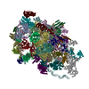
| Title | Structure of 70S ribosome in the 100S ribosome in the hibernation stage | |||||||||
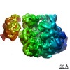
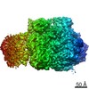
 Map data Map data | The density map of 70S ribosome with part of its partner in the 100S ribosome in hibernation stage. | |||||||||
 Sample Sample |
| |||||||||
 Keywords Keywords | cryoelectron microscopy / ribosomal protein | |||||||||
| Biological species |  | |||||||||
| Method | single particle reconstruction / cryo EM / Resolution: 18.0 Å | |||||||||
 Authors Authors | Kato T / Yoshida H / Miyata T / Maki Y / Wada A / Namba K | |||||||||
 Citation Citation | Journal: Structure / Year: 2010 Title: Structure of the 100S ribosome in the hibernation stage revealed by electron cryomicroscopy. Authors: Takayuki Kato / Hideji Yoshida / Tomoko Miyata / Yasushi Maki / Akira Wada / Keiichi Namba /  Abstract: In the stationary growth phase of bacteria, protein biosynthesis on ribosomes is suppressed, and the ribosomes are preserved in the cell by the formation of the 100S ribosome. The 100S ribosome is a ...In the stationary growth phase of bacteria, protein biosynthesis on ribosomes is suppressed, and the ribosomes are preserved in the cell by the formation of the 100S ribosome. The 100S ribosome is a dimer of the 70S ribosome and is formed by the binding of the ribosome modulation factor and the hibernation promoting factor. However, the binding mode between the two 70S ribosomes and the mechanism of complex formation are still poorly understood. Here, we report the structure of the 100S ribosome by electron cryomicroscopy and single-particle image analysis. The 100S ribosome purified from the cell in the stationary growth phase is composed of two transfer RNA-free 70S ribosomes, has two-fold symmetry, and is formed through interactions between their 30S subunits, where interactions between small subunit proteins, S2, S3 and S5, appear to be critical for the dimerization. | |||||||||
| History |
|
- Structure visualization
| Movie |
 Movie viewer Movie viewer |
|---|---|
| Structure viewer | EM map: SurfViewMolmilJmol/JSmol |
| Supplemental images |


- Downloads & links
-EMDB archive
| Map data | emd_5174.map.gz | 59.5 MB | EMDB map data format | |
|---|---|---|---|---|
| Header (meta data) | emd-5174-v30.xmlemd-5174.xml | 10 KB 10 KB | Display Display | EMDB header |
| Images | 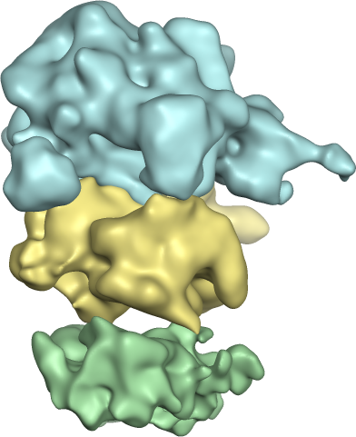 emd_5174.png emd_5174.png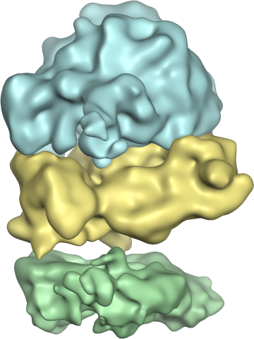 emd_5174_1.png emd_5174_1.png | 132 KB 139.9 KB | ||
| Archive directory |  http://ftp.pdbj.org/pub/emdb/structures/EMD-5174ftp://ftp.pdbj.org/pub/emdb/structures/EMD-5174 http://ftp.pdbj.org/pub/emdb/structures/EMD-5174ftp://ftp.pdbj.org/pub/emdb/structures/EMD-5174 | HTTPS FTP |
-Related structure data
| Similar structure data |
|---|

-Links
| EMDB pages | EMDB (EBI/PDBe) / EMDataResource |
|---|---|
| Related items in Molecule of the Month |

-Map
| File | Download / File: emd_5174.map.gz / Format: CCP4 / Size: 62.5 MB / Type: IMAGE STORED AS FLOATING POINT NUMBER (4 BYTES) | ||||||||||||||||||||||||||||||||||||||||||||||||||||||||||||||||||||
|---|---|---|---|---|---|---|---|---|---|---|---|---|---|---|---|---|---|---|---|---|---|---|---|---|---|---|---|---|---|---|---|---|---|---|---|---|---|---|---|---|---|---|---|---|---|---|---|---|---|---|---|---|---|---|---|---|---|---|---|---|---|---|---|---|---|---|---|---|---|
| Annotation | The density map of 70S ribosome with part of its partner in the 100S ribosome in hibernation stage. | ||||||||||||||||||||||||||||||||||||||||||||||||||||||||||||||||||||
| Projections & slices | Image control
Images are generated by Spider. | ||||||||||||||||||||||||||||||||||||||||||||||||||||||||||||||||||||
| Voxel size | X=Y=Z: 1.69 Å | ||||||||||||||||||||||||||||||||||||||||||||||||||||||||||||||||||||
| Density |
| ||||||||||||||||||||||||||||||||||||||||||||||||||||||||||||||||||||
| Symmetry | Space group: 1 | ||||||||||||||||||||||||||||||||||||||||||||||||||||||||||||||||||||
| Details | EMDB XML:
CCP4 map header:
| ||||||||||||||||||||||||||||||||||||||||||||||||||||||||||||||||||||
 Z (Sec.)
 Y (Row.)
 X (Col.)


-Supplemental data
- Sample components
-Entire : E. coli 100S ribosome
| Entire | Name: E. coli 100S ribosome |
|---|---|
| Components |
|
-Supramolecule #1000: E. coli 100S ribosome
| Supramolecule | Name: E. coli 100S ribosome / type: sample / ID: 1000 Details: The 100S ribosome was fixed by GraFix with glutaraldehyde. Oligomeric state: dimer of 70S ribosome / Number unique components: 1 |
|---|
-Supramolecule #1: 100S ribosome
| Supramolecule | Name: 100S ribosome / type: complex / ID: 1 / Recombinant expression: No / Database: NCBI / Ribosome-details: ribosome-prokaryote: ALL |
|---|---|
| Source (natural) | Organism: |
-Experimental details
-Structure determination
| Method | cryo EM |
|---|---|
 Processing Processing | single particle reconstruction |
| Aggregation state | particle |
-Sample preparation
| Buffer | pH: 7.6 / Details: 20 mM HEPES-KCL, 77 mM KCl, 15 mM (CH3COO)2Mg |
|---|---|
| Grid | Details: Quantifoil 0.6/1 on 200 mesh molybdenum grid |
| Vitrification | Cryogen name: ETHANE / Chamber humidity: 90 % / Chamber temperature: 100 K / Instrument: OTHER / Details: Vitrification instrument: FEI Vitrobot / Method: Blot for 7 seconds before plunging |
- Electron microscopy
| Microscope | JEOL 3200FSC |
|---|---|
| Temperature | Min: 50 K / Max: 70 K |
| Specialist optics | Energy filter - Name: JEOL Omega filter / Energy filter - Lower energy threshold: 0.0 eV / Energy filter - Upper energy threshold: 10.0 eV |
| Date | Feb 25, 2009 |
| Image recording | Category: CCD / Film or detector model: TVIPS TEMCAM-F415 (4k x 4k) / Digitization - Sampling interval: 15 µm / Number real images: 300 / Average electron dose: 20 e/Å2 / Bits/pixel: 16 |
| Electron beam | Acceleration voltage: 200 kV / Electron source:  FIELD EMISSION GUN FIELD EMISSION GUN |
| Electron optics | Calibrated magnification: 88760 / Illumination mode: FLOOD BEAM / Imaging mode: BRIGHT FIELD / Cs: 1.6 mm / Nominal defocus max: 3.5 µm / Nominal defocus min: 1.25 µm / Nominal magnification: 50000 |
| Sample stage | Specimen holder: Top entry liquid helium-cooled cryo specimen holder Specimen holder model: JEOL |
-Image processing
| Details | Each of the two 70S ribosome particle images with part of the dimer pair was extracted in a box with circler mask from 100S ribosome particle image. |
|---|---|
| CTF correction | Details: Each particle |
| Final reconstruction | Algorithm: OTHER / Resolution.type: BY AUTHOR / Resolution: 18.0 Å / Resolution method: FSC 0.5 CUT-OFF / Software - Name: SPIDER / Number images used: 17461 |
| Final two d classification | Number classes: 552 |
