Movie
Movie Controller


+ Open data

- Basic information
| Entry |  | |||||||||||||||||||||
|---|---|---|---|---|---|---|---|---|---|---|---|---|---|---|---|---|---|---|---|---|---|---|
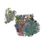
| Title | 30S mRNA delivery complex (closed head) focused body map | |||||||||||||||||||||
 Map data Map data | ||||||||||||||||||||||
 Sample Sample |
| |||||||||||||||||||||
 Keywords Keywords | Transcription / translation / coupling / RIBOSOME | |||||||||||||||||||||
| Biological species |  | |||||||||||||||||||||
| Method | single particle reconstruction / cryo EM / Resolution: 2.7 Å | |||||||||||||||||||||
 Authors Authors | Rahil H / Weixlbaumer A / Webster MW | |||||||||||||||||||||
| Funding support | European Union,  France, France,  United States, United States,  United Kingdom, 6 items United Kingdom, 6 items
| |||||||||||||||||||||
 Citation Citation | Journal: Science / Year: 2024 Title: Molecular basis of mRNA delivery to the bacterial ribosome. Authors: Michael W Webster / Adrien Chauvier / Huma Rahil / Andrea Graziadei / Kristine Charles / Nataliya Miropolskaya / Maria Takacs / Charlotte Saint-André / Juri Rappsilber / Nils G Walter / Albert Weixlbaumer /  Abstract: Protein synthesis begins with the formation of a ribosome-messenger RNA (mRNA) complex. In bacteria, the small ribosomal subunit (30) is recruited to many mRNAs through base pairing with the Shine- ...Protein synthesis begins with the formation of a ribosome-messenger RNA (mRNA) complex. In bacteria, the small ribosomal subunit (30) is recruited to many mRNAs through base pairing with the Shine-Dalgarno (SD) sequence and RNA binding by ribosomal protein bS1. Translation can initiate on nascent mRNAs, and RNA polymerase (RNAP) can promote the recruitment of the pioneering 30. Here, we examined 30 recruitment to nascent mRNAs using cryo-electron microscopy, single-molecule fluorescence colocalization, and in-cell cross-linking mass spectrometry. We show that bS1 delivers the mRNA to the ribosome for SD duplex formation and 30 activation. Additionally, bS1 and RNAP stimulate translation initiation. Our work provides a mechanistic framework for how the SD duplex, ribosomal proteins, and RNAP cooperate in 30 recruitment to mRNAs and establish transcription-translation coupling. | |||||||||||||||||||||
| History |
|
- Structure visualization
| Supplemental images |
|---|

- Downloads & links
-EMDB archive
| Map data | emd_51589.map.gz | 778.7 MB |  EMDB map data format EMDB map data format | |
|---|---|---|---|---|
| Header (meta data) | emd-51589-v30.xmlemd-51589.xml | 18.7 KB 18.7 KB | Display Display | EMDB header |
| FSC (resolution estimation) | emd_51589_fsc.xml | 19.8 KB | Display | FSC data file |
| Images |  emd_51589.png emd_51589.png | 60.2 KB | ||
| Masks | emd_51589_msk_1.map | 824 MB | Mask map | |
| Filedesc metadata | emd-51589.cif.gz | 4.2 KB | ||
| Others | emd_51589_additional_1.map.gzemd_51589_half_map_1.map.gzemd_51589_half_map_2.map.gz | 413.5 MB 765.7 MB 765.7 MB | ||
| Archive directory |  http://ftp.pdbj.org/pub/emdb/structures/EMD-51589ftp://ftp.pdbj.org/pub/emdb/structures/EMD-51589 http://ftp.pdbj.org/pub/emdb/structures/EMD-51589ftp://ftp.pdbj.org/pub/emdb/structures/EMD-51589 | HTTPS FTP |
-Validation report
| Summary document | emd_51589_validation.pdf.gz | 1.1 MB | Display | EMDB validaton report |
|---|---|---|---|---|
| Full document | emd_51589_full_validation.pdf.gz | 1.1 MB | Display | |
| Data in XML | emd_51589_validation.xml.gz | 28.3 KB | Display | |
| Data in CIF | emd_51589_validation.cif.gz | 37.4 KB | Display | |
| Arichive directory | https://ftp.pdbj.org/pub/emdb/validation_reports/EMD-51589ftp://ftp.pdbj.org/pub/emdb/validation_reports/EMD-51589 | HTTPS FTP |
-Related structure data
| Related structure data |  9gr1C  9gupC  9guqC  9gurC  9gusC  9gutC  9guuC  9guvC  9guwC  9guxC C: citing same article ( |
|---|


-Links
| EMDB pages | EMDB (EBI/PDBe) / EMDataResource |
|---|
-Map
| File | Download / File: emd_51589.map.gz / Format: CCP4 / Size: 824 MB / Type: IMAGE STORED AS FLOATING POINT NUMBER (4 BYTES) | ||||||||||||||||||||||||||||||||||||
|---|---|---|---|---|---|---|---|---|---|---|---|---|---|---|---|---|---|---|---|---|---|---|---|---|---|---|---|---|---|---|---|---|---|---|---|---|---|
| Projections & slices | Image control
Images are generated by Spider. | ||||||||||||||||||||||||||||||||||||
| Voxel size | X=Y=Z: 0.84 Å | ||||||||||||||||||||||||||||||||||||
| Density |
| ||||||||||||||||||||||||||||||||||||
| Symmetry | Space group: 1 | ||||||||||||||||||||||||||||||||||||
| Details | EMDB XML:
|
 Z (Sec.)
 Y (Row.)
 X (Col.)


-Supplemental data
-Mask #1
| File | emd_51589_msk_1.map | ||||||||||||
|---|---|---|---|---|---|---|---|---|---|---|---|---|---|
| Projections & Slices |
| ||||||||||||
| Density Histograms |


-Additional map: #1
| File | emd_51589_additional_1.map | ||||||||||||
|---|---|---|---|---|---|---|---|---|---|---|---|---|---|
| Projections & Slices |
| ||||||||||||
| Density Histograms |


-Half map: #1
| File | emd_51589_half_map_1.map | ||||||||||||
|---|---|---|---|---|---|---|---|---|---|---|---|---|---|
| Projections & Slices |
| ||||||||||||
| Density Histograms |


-Half map: #2
| File | emd_51589_half_map_2.map | ||||||||||||
|---|---|---|---|---|---|---|---|---|---|---|---|---|---|
| Projections & Slices |
| ||||||||||||
| Density Histograms |


- Sample components
-Entire : 30S ribosomal subunit
| Entire | Name: 30S ribosomal subunit |
|---|---|
| Components |
|
-Supramolecule #1: 30S ribosomal subunit
| Supramolecule | Name: 30S ribosomal subunit / type: complex / ID: 1 / Parent: 0 / Macromolecule list: #1-#24 |
|---|---|
| Source (natural) | Organism: |
-Experimental details
-Structure determination
| Method | cryo EM |
|---|---|
 Processing Processing | single particle reconstruction |
| Aggregation state | particle |
-Sample preparation
| Buffer | pH: 7.4 |
|---|---|
| Vitrification | Cryogen name: ETHANE |
- Electron microscopy
| Microscope | FEI TITAN KRIOS |
|---|---|
| Image recording | Film or detector model: GATAN K2 QUANTUM (4k x 4k) / Average electron dose: 49.95 e/Å2 |
| Electron beam | Acceleration voltage: 300 kV / Electron source:  FIELD EMISSION GUN FIELD EMISSION GUN |
| Electron optics | Illumination mode: FLOOD BEAM / Imaging mode: BRIGHT FIELD / Nominal defocus max: 2.0 µm / Nominal defocus min: 0.8 µm |
| Experimental equipment |  Model: Titan Krios / Image courtesy: FEI Company |