farnesyl-diphosphate kinase / farnesyl diphosphate kinase activity / succinyl-CoA binding / ITP biosynthetic process / isoprenoid metabolic process / Hydrolases; Acting on ester bonds; Site specific endodeoxyribonucleases: cleavage is not sequence specific (deleted sub-subclass) / phosphotransferase activity, phosphate group as acceptor / DNA nuclease activity / acetyl-CoA catabolic process / nucleoside triphosphate biosynthetic process ...farnesyl-diphosphate kinase / farnesyl diphosphate kinase activity / succinyl-CoA binding / ITP biosynthetic process / isoprenoid metabolic process / Hydrolases; Acting on ester bonds; Site specific endodeoxyribonucleases: cleavage is not sequence specific (deleted sub-subclass) / phosphotransferase activity, phosphate group as acceptor / DNA nuclease activity / acetyl-CoA catabolic process / nucleoside triphosphate biosynthetic process / acetyl-CoA binding / nucleoside diphosphate metabolic process / Ribavirin ADME / coenzyme A binding / protein histidine kinase activity / regulation of fatty acid biosynthetic process / apoptotic DNA fragmentation / nucleoside-diphosphate kinase / 3'-5'-DNA exonuclease activity / Interconversion of nucleotide di- and triphosphates / UTP biosynthetic process / CTP biosynthetic process / protein hexamerization / Azathioprine ADME / DNA catabolic process / nucleoside diphosphate kinase activity / GTP biosynthetic process / Hydrolases; Acting on ester bonds; Exodeoxyribonucleases producing 5'-phosphomonoesters / histidine kinase / ribosomal small subunit binding / lactation / positive regulation of epithelial cell proliferation / DNA endonuclease activity / ADP binding / ruffle membrane / endocytosis / kinase activity / GDP binding / nervous system development / regulation of apoptotic process / early endosome / cell differentiation / non-specific serine/threonine protein kinase / negative regulation of cell population proliferation / magnesium ion binding / DNA binding / RNA binding / extracellular exosome / ATP binding / membrane / identical protein binding / nucleus / cytoplasm / cytosol Similarity search - Function
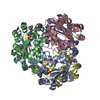
Journal: Nat Chem / Year: 2025 Title: Nucleoside diphosphate kinase A (NME1) catalyses its own oligophosphorylation. Authors: Arif Celik / Felix Schöpf / Christian E Stieger / Sarah Lampe / Björn Hanf / Jeremy A M Morgan / Max Ruwolt / Fan Liu / Christian P R Hackenberger / Daniel Roderer / Dorothea Fiedler / Abstract: Protein phosphorylation is a central signalling mechanism in eukaryotic cells. The scope of this post-translational modification includes protein pyro- and polyphosphorylation. Here we report the ...Protein phosphorylation is a central signalling mechanism in eukaryotic cells. The scope of this post-translational modification includes protein pyro- and polyphosphorylation. Here we report the discovery of another mode of phosphorylation: protein oligophosphorylation. Using site-specifically phosphorylated and pyrophosphorylated nucleoside diphosphate kinase A (NME1), the effects of these modifications on enzyme activity were investigated. Phosphorylation, and more so pyrophosphorylation, on Thr94 reduced the nucleoside diphosphate kinase activity. Nevertheless, both phosphoprotein and pyrophosphoprotein catalysed their own oligophosphorylation-up to the formation of a hexaphosphate chain-using ATP as a cofactor. Oligophosphorylation was critically dependent on the catalytic histidine residue His118, and cryogenic electron microscopy analysis of the modified proteins suggests an intramolecular phosphoryl transfer mechanism. Oligophosphorylation of NME1 in biochemical samples, and in cell lysates, was further confirmed using mass spectrometry, and was found to promote a new set of protein interactions. Our results highlight the complex nature of phosphoregulation, and the methods described here provide the opportunity to investigate the impact of this unusual modification in the future.
Cryogen name: ETHANE / Chamber humidity: 100 % / Chamber temperature: 285 K / Instrument: FEI VITROBOT MARK IV
-
Electron microscopy
Microscope
TFS KRIOS
Specialist optics
Energy filter - Name: GIF Bioquantum / Energy filter - Slit width: 20 eV
Image recording
Film or detector model: GATAN K3 BIOQUANTUM (6k x 4k) / Number grids imaged: 1 / Number real images: 4639 / Average exposure time: 2.0 sec. / Average electron dose: 44.6 e/Å2
Electron beam
Acceleration voltage: 300 kV / Electron source: FIELD EMISSION GUN
In the structure databanks used in Yorodumi, some data are registered as the other names, "COVID-19 virus" and "2019-nCoV". Here are the details of the virus and the list of structure data.
Jan 31, 2019. EMDB accession codes are about to change! (news from PDBe EMDB page)
EMDB accession codes are about to change! (news from PDBe EMDB page)
The allocation of 4 digits for EMDB accession codes will soon come to an end. Whilst these codes will remain in use, new EMDB accession codes will include an additional digit and will expand incrementally as the available range of codes is exhausted. The current 4-digit format prefixed with “EMD-” (i.e. EMD-XXXX) will advance to a 5-digit format (i.e. EMD-XXXXX), and so on. It is currently estimated that the 4-digit codes will be depleted around Spring 2019, at which point the 5-digit format will come into force.
The EM Navigator/Yorodumi systems omit the EMD- prefix.
Related info.:Q: What is EMD? / ID/Accession-code notation in Yorodumi/EM Navigator
Yorodumi is a browser for structure data from EMDB, PDB, SASBDB, etc.
This page is also the successor to EM Navigator detail page, and also detail information page/front-end page for Omokage search.
The word "yorodu" (or yorozu) is an old Japanese word meaning "ten thousand". "mi" (miru) is to see.
Related info.:EMDB / PDB / SASBDB / Comparison of 3 databanks / Yorodumi Search / Aug 31, 2016. New EM Navigator & Yorodumi / Yorodumi Papers / Jmol/JSmol / Function and homology information / Changes in new EM Navigator and Yorodumi
 Movie
Movie Controller


 Open data
 Basic information
 Map data
 Sample
 Keywords
 Function and homology information
 Homo sapiens (human)
 Authors
 Germany, 1 items
  Citation
 Structure visualization
 Downloads & links
 emd_51250.png
 http://ftp.pdbj.org/pub/emdb/structures/EMD-51250


 Z (Sec.)
 Y (Row.)
 X (Col.)


 Sample components
 Processing
 Electron microscopy
 FIELD EMISSION GUN