 Movie
Movie Controller


+ Open data

- Basic information
| Entry |  | |||||||||
|---|---|---|---|---|---|---|---|---|---|---|
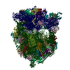
| Title | Untethered Cytoplasmic Fission Yeast Ribosome (S. pombe) | |||||||||
 Map data Map data | Untethered Cytosolic Ribosome of S. pombe generated by M | |||||||||
 Sample Sample |
| |||||||||
 Keywords Keywords | Hibernating / Yeast / In Situ / RACK1 / cytosolic / RIBOSOME / untethered | |||||||||
| Biological species |  | |||||||||
| Method | subtomogram averaging / cryo EM / Resolution: 14.2 Å | |||||||||
 Authors Authors | Rosa H / Gemin O / Gluc M / Mattei S / Jomaa A | |||||||||
| Funding support | 1 items
| |||||||||
 Citation Citation | Journal: Nat Commun / Year: 2024 Title: Ribosomes hibernate on mitochondria during cellular stress. Authors: Olivier Gemin / Maciej Gluc / Higor Rosa / Michael Purdy / Moritz Niemann / Yelena Peskova / Simone Mattei / Ahmad Jomaa /   Abstract: Cell survival under nutrient-deprived conditions relies on cells' ability to adapt their organelles and rewire their metabolic pathways. In yeast, glucose depletion induces a stress response mediated ...Cell survival under nutrient-deprived conditions relies on cells' ability to adapt their organelles and rewire their metabolic pathways. In yeast, glucose depletion induces a stress response mediated by mitochondrial fragmentation and sequestration of cytosolic ribosomes on mitochondria. This cellular adaptation promotes survival under harsh environmental conditions; however, the underlying mechanism of this response remains unknown. Here, we demonstrate that upon glucose depletion protein synthesis is halted. Cryo-electron microscopy structure of the ribosomes show that they are devoid of both tRNA and mRNA, and a subset of the particles depicted a conformational change in rRNA H69 that could prevent tRNA binding. Our in situ structural analyses reveal that the hibernating ribosomes tether to fragmented mitochondria and establish eukaryotic-specific, higher-order storage structures by assembling into oligomeric arrays on the mitochondrial surface. Notably, we show that hibernating ribosomes exclusively bind to the outer mitochondrial membrane via the small ribosomal subunit during cellular stress. We identify the ribosomal protein Cpc2/RACK1 as the molecule mediating ribosomal tethering to mitochondria. This study unveils the molecular mechanism connecting mitochondrial stress with the shutdown of protein synthesis and broadens our understanding of cellular responses to nutrient scarcity and cell quiescence. | |||||||||
| History |
|
- Structure visualization
| Supplemental images |
|---|

- Downloads & links
-EMDB archive
| Map data | emd_51030.map.gz | 13 MB |  EMDB map data format EMDB map data format | |
|---|---|---|---|---|
| Header (meta data) | emd-51030-v30.xmlemd-51030.xml | 13.4 KB 13.4 KB | Display Display | EMDB header |
| FSC (resolution estimation) | emd_51030_fsc.xml | 5.5 KB | Display | FSC data file |
| Images |  emd_51030.png emd_51030.png | 124.7 KB | ||
| Filedesc metadata | emd-51030.cif.gz | 4.2 KB | ||
| Others | emd_51030_half_map_1.map.gzemd_51030_half_map_2.map.gz | 7.1 MB 7.1 MB | ||
| Archive directory |  http://ftp.pdbj.org/pub/emdb/structures/EMD-51030ftp://ftp.pdbj.org/pub/emdb/structures/EMD-51030 http://ftp.pdbj.org/pub/emdb/structures/EMD-51030ftp://ftp.pdbj.org/pub/emdb/structures/EMD-51030 | HTTPS FTP |
-Validation report
| Summary document | emd_51030_validation.pdf.gz | 829.5 KB | Display | EMDB validaton report |
|---|---|---|---|---|
| Full document | emd_51030_full_validation.pdf.gz | 829.1 KB | Display | |
| Data in XML | emd_51030_validation.xml.gz | 11.1 KB | Display | |
| Data in CIF | emd_51030_validation.cif.gz | 15.2 KB | Display | |
| Arichive directory | https://ftp.pdbj.org/pub/emdb/validation_reports/EMD-51030ftp://ftp.pdbj.org/pub/emdb/validation_reports/EMD-51030 | HTTPS FTP |
-Related structure data


-Links
| EMDB pages | EMDB (EBI/PDBe) / EMDataResource |
|---|
-Map
| File | Download / File: emd_51030.map.gz / Format: CCP4 / Size: 13.9 MB / Type: IMAGE STORED AS FLOATING POINT NUMBER (4 BYTES) | ||||||||||||||||||||||||||||||||||||
|---|---|---|---|---|---|---|---|---|---|---|---|---|---|---|---|---|---|---|---|---|---|---|---|---|---|---|---|---|---|---|---|---|---|---|---|---|---|
| Annotation | Untethered Cytosolic Ribosome of S. pombe generated by M | ||||||||||||||||||||||||||||||||||||
| Projections & slices | Image control
Images are generated by Spider. | ||||||||||||||||||||||||||||||||||||
| Voxel size | X=Y=Z: 4.15 Å | ||||||||||||||||||||||||||||||||||||
| Density |
| ||||||||||||||||||||||||||||||||||||
| Symmetry | Space group: 1 | ||||||||||||||||||||||||||||||||||||
| Details | EMDB XML:
|
 Z (Sec.)
 Y (Row.)
 X (Col.)


-Supplemental data
-Half map: Untethered Cytosolic Ribosome of S. pombe Half-Map 2 generated by M
| File | emd_51030_half_map_1.map | ||||||||||||
|---|---|---|---|---|---|---|---|---|---|---|---|---|---|
| Annotation | Untethered Cytosolic Ribosome of S. pombe Half-Map 2 generated by M | ||||||||||||
| Projections & Slices |
| ||||||||||||
| Density Histograms |


-Half map: Untethered Cytosolic Ribosome of S. pombe Half-Map 1 generated by M
| File | emd_51030_half_map_2.map | ||||||||||||
|---|---|---|---|---|---|---|---|---|---|---|---|---|---|
| Annotation | Untethered Cytosolic Ribosome of S. pombe Half-Map 1 generated by M | ||||||||||||
| Projections & Slices |
| ||||||||||||
| Density Histograms |


- Sample components
-Entire : Cytoplasmic Ribosome Untethered to the Outer Mitochondrial Membrane
| Entire | Name: Cytoplasmic Ribosome Untethered to the Outer Mitochondrial Membrane |
|---|---|
| Components |
|
-Supramolecule #1: Cytoplasmic Ribosome Untethered to the Outer Mitochondrial Membrane
| Supramolecule | Name: Cytoplasmic Ribosome Untethered to the Outer Mitochondrial Membrane type: complex / ID: 1 / Parent: 0 |
|---|---|
| Source (natural) | Organism: |
-Experimental details
-Structure determination
| Method | cryo EM |
|---|---|
 Processing Processing | subtomogram averaging |
| Aggregation state | cell |
-Sample preparation
| Buffer | pH: 5.5 |
|---|---|
| Vitrification | Cryogen name: ETHANE / Chamber humidity: 100 % / Chamber temperature: 300 K / Instrument: LEICA EM GP |
- Electron microscopy
| Microscope | FEI TITAN KRIOS |
|---|---|
| Specialist optics | Energy filter - Name: GIF Bioquantum / Energy filter - Slit width: 30 eV |
| Image recording | Film or detector model: GATAN K3 BIOQUANTUM (6k x 4k) / Average electron dose: 2.23 e/Å2 |
| Electron beam | Acceleration voltage: 300 kV / Electron source:  FIELD EMISSION GUN FIELD EMISSION GUN |
| Electron optics | C2 aperture diameter: 50.0 µm / Illumination mode: FLOOD BEAM / Imaging mode: BRIGHT FIELD / Cs: 2.7 mm / Nominal defocus max: 4.0 µm / Nominal defocus min: 2.0 µm / Nominal magnification: 42000 |
| Sample stage | Specimen holder model: FEI TITAN KRIOS AUTOGRID HOLDER / Cooling holder cryogen: NITROGEN |
| Experimental equipment |  Model: Titan Krios / Image courtesy: FEI Company |
-Image processing
| Final reconstruction | Applied symmetry - Point group: C1 (asymmetric) / Resolution.type: BY AUTHOR / Resolution: 14.2 Å / Resolution method: FSC 0.143 CUT-OFF / Software - Name: Warp (ver. 1.09) / Number subtomograms used: 2415 |
|---|---|
| Extraction | Number tomograms: 53 / Number images used: 12048 |
| Final angle assignment | Type: MAXIMUM LIKELIHOOD / Details: Relion (Refine) + M (PPoses) |
| FSC plot (resolution estimation) |  |
