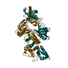
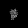
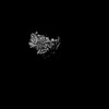
Journal: Sci Adv / Year: 2025 Title: Single-stranded DNA binding to the transcription factor PafBC triggers the mycobacterial DNA damage response. Authors: Charlotte M Schilling / Rafal Zdanowicz / Julius Rabl / Andreas U Müller / Daniel Boehringer / Rudi Glockshuber / Eilika Weber-Ban / Abstract: The DNA damage response in mycobacteria is controlled by the heterodimeric transcription factor PafBC, a member of the WYL domain-containing protein family. It has been shown that PafBC induces ...The DNA damage response in mycobacteria is controlled by the heterodimeric transcription factor PafBC, a member of the WYL domain-containing protein family. It has been shown that PafBC induces transcription of its regulon by reprogramming the housekeeping RNA polymerase holoenzyme to recognize PafBC-dependent promoters through sigma adaptation. However, the mechanism by which DNA damage is sensed and translated into PafBC activation has remained unclear. Here, we demonstrate that the binding of single-stranded DNA (ssDNA) to the WYL domains of PafBC activates the transcription factor. Our cryo-electron microscopy structure of full-length PafBC in its active conformation, bound to the transcription initiation complex, reveals a previously unknown mode of interaction between the ssDNA and the WYL domains. Using biochemical experiments, we show that short ssDNA fragments bind to PafBC dynamically, resulting in deactivation as ssDNA levels decrease postrepair. Our findings shed light on the mechanism linking DNA damage to PafBC activation and expand our understanding of WYL domain-containing proteins.
In the structure databanks used in Yorodumi, some data are registered as the other names, "COVID-19 virus" and "2019-nCoV". Here are the details of the virus and the list of structure data.
Jan 31, 2019. EMDB accession codes are about to change! (news from PDBe EMDB page)
EMDB accession codes are about to change! (news from PDBe EMDB page)
The allocation of 4 digits for EMDB accession codes will soon come to an end. Whilst these codes will remain in use, new EMDB accession codes will include an additional digit and will expand incrementally as the available range of codes is exhausted. The current 4-digit format prefixed with “EMD-” (i.e. EMD-XXXX) will advance to a 5-digit format (i.e. EMD-XXXXX), and so on. It is currently estimated that the 4-digit codes will be depleted around Spring 2019, at which point the 5-digit format will come into force.
The EM Navigator/Yorodumi systems omit the EMD- prefix.
Related info.:Q: What is EMD? / ID/Accession-code notation in Yorodumi/EM Navigator
Yorodumi is a browser for structure data from EMDB, PDB, SASBDB, etc.
This page is also the successor to EM Navigator detail page, and also detail information page/front-end page for Omokage search.
The word "yorodu" (or yorozu) is an old Japanese word meaning "ten thousand". "mi" (miru) is to see.
Related info.:EMDB / PDB / SASBDB / Comparison of 3 databanks / Yorodumi Search / Aug 31, 2016. New EM Navigator & Yorodumi / Yorodumi Papers / Jmol/JSmol / Function and homology information / Changes in new EM Navigator and Yorodumi
 Movie
Movie Controller

 Yorodumi
 Open data
 Basic information
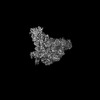
 Map data
 Sample
 Keywords
 Function and homology information
 Mycolicibacterium smegmatis MC2 155 (bacteria)
 Authors
 Switzerland, 1 items
  Citation
 Structure visualization
 Downloads & links
 emd_50591.png
 http://ftp.pdbj.org/pub/emdb/structures/EMD-50591
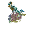


 X (Sec.)
 Y (Row.)
 Z (Col.)


 Sample components
 Processing
 Electron microscopy
 FIELD EMISSION GUN
