 Movie
Movie Controller

[English] 日本語
 Yorodumi
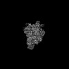
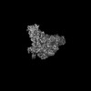
- PDB-9fnd: Transcriptional activator PafBC bound to mycobacterial RNA polymerase -
+ Open data

- Basic information
| Entry | Database: PDB / ID: 9fnd | ||||||
|---|---|---|---|---|---|---|---|
| Title | Transcriptional activator PafBC bound to mycobacterial RNA polymerase | ||||||
 Components Components |
| ||||||
 Keywords Keywords | TRANSCRIPTION / PafBC / sigma adaptation / WYL domain / transcriptional activator | ||||||
| Function / homology |  Function and homology information Function and homology informationProtein PafC / PafC, HTH domain / PafC helix-turn-helix domain / : / WCX domain / : / WYL domain profile. / WYL domain / WYL domain Similarity search - Domain/homology | ||||||
| Biological species |  Mycolicibacterium smegmatis MC2 155 (bacteria) Mycolicibacterium smegmatis MC2 155 (bacteria) | ||||||
| Method | ELECTRON MICROSCOPY / single particle reconstruction / cryo EM / Resolution: 4 Å | ||||||
 Authors Authors | Zdanowicz, R. / Schilling, C.M. / Rabl, J. / Mueller, A.U. / Boehringer, D. / Glockshuber, R. / Weber-Ban, E. | ||||||
| Funding support |  Switzerland, 1items Switzerland, 1items
| ||||||
 Citation Citation | Journal: Sci Adv / Year: 2025 Title: Single-stranded DNA binding to the transcription factor PafBC triggers the mycobacterial DNA damage response. Authors: Charlotte M Schilling / Rafal Zdanowicz / Julius Rabl / Andreas U Müller / Daniel Boehringer / Rudi Glockshuber / Eilika Weber-Ban / Abstract: The DNA damage response in mycobacteria is controlled by the heterodimeric transcription factor PafBC, a member of the WYL domain-containing protein family. It has been shown that PafBC induces ...The DNA damage response in mycobacteria is controlled by the heterodimeric transcription factor PafBC, a member of the WYL domain-containing protein family. It has been shown that PafBC induces transcription of its regulon by reprogramming the housekeeping RNA polymerase holoenzyme to recognize PafBC-dependent promoters through sigma adaptation. However, the mechanism by which DNA damage is sensed and translated into PafBC activation has remained unclear. Here, we demonstrate that the binding of single-stranded DNA (ssDNA) to the WYL domains of PafBC activates the transcription factor. Our cryo-electron microscopy structure of full-length PafBC in its active conformation, bound to the transcription initiation complex, reveals a previously unknown mode of interaction between the ssDNA and the WYL domains. Using biochemical experiments, we show that short ssDNA fragments bind to PafBC dynamically, resulting in deactivation as ssDNA levels decrease postrepair. Our findings shed light on the mechanism linking DNA damage to PafBC activation and expand our understanding of WYL domain-containing proteins. | ||||||
| History |
|
- Structure visualization
| Structure viewer | Molecule: MolmilJmol/JSmol |
|---|
- Downloads & links
-Download
| PDBx/mmCIF format | 9fnd.cif.gz | 118.3 KB | Display | PDBx/mmCIF format |
|---|---|---|---|---|
| PDB format | pdb9fnd.ent.gz | 90.3 KB | Display | PDB format |
| PDBx/mmJSON format | 9fnd.json.gz | Tree view | PDBx/mmJSON format | |
| Others |  Other downloads Other downloads |
-Validation report
| Arichive directory | https://data.pdbj.org/pub/pdb/validation_reports/fn/9fndftp://data.pdbj.org/pub/pdb/validation_reports/fn/9fnd | HTTPS FTP |
|---|
-Related structure data
| Related structure data | 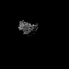 50590MC 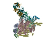 9fneC M: map data used to model this data C: citing same article ( |
|---|---|
| Similar structure data |
-Links
 PDBj
PDBj- Assembly
| Deposited unit | 
|
|---|---|
| 1 |
|
-Components
| #1: Protein | Mass: 36207.555 Da / Num. of mol.: 1 Source method: isolated from a genetically manipulated source Source: (gene. exp.) Mycolicibacterium smegmatis MC2 155 (bacteria)Gene: pafB, MSMEI_3799 / Production host: |
|---|---|
| #2: Protein | Mass: 34044.402 Da / Num. of mol.: 1 Source method: isolated from a genetically manipulated source Source: (gene. exp.) Mycolicibacterium smegmatis MC2 155 (bacteria)Gene: MSMEG_3888 / Production host: |
| Has protein modification | N |
-Experimental details
-Experiment
| Experiment | Method: ELECTRON MICROSCOPY |
|---|---|
| EM experiment | Aggregation state: PARTICLE / 3D reconstruction method: single particle reconstruction |
- Sample preparation
| Component |
| ||||||||||||||||||||||||||||||
|---|---|---|---|---|---|---|---|---|---|---|---|---|---|---|---|---|---|---|---|---|---|---|---|---|---|---|---|---|---|---|---|
| Molecular weight |
| ||||||||||||||||||||||||||||||
| Source (natural) |
| ||||||||||||||||||||||||||||||
| Source (recombinant) |
| ||||||||||||||||||||||||||||||
| Buffer solution | pH: 8 | ||||||||||||||||||||||||||||||
| Specimen | Embedding applied: NO / Shadowing applied: NO / Staining applied: NO / Vitrification applied: YES | ||||||||||||||||||||||||||||||
| Specimen support | Grid material: COPPER / Grid mesh size: 300 divisions/in. / Grid type: Quantifoil R2/2 | ||||||||||||||||||||||||||||||
| Vitrification | Instrument: FEI VITROBOT MARK IV / Cryogen name: ETHANE-PROPANE / Humidity: 95 % / Chamber temperature: 277.15 K |
- Electron microscopy imaging
| Experimental equipment |  Model: Titan Krios / Image courtesy: FEI Company |
|---|---|
| Microscopy | Model: FEI TITAN KRIOS |
| Electron gun | Electron source:  FIELD EMISSION GUN / Accelerating voltage: 300 kV / Illumination mode: FLOOD BEAM FIELD EMISSION GUN / Accelerating voltage: 300 kV / Illumination mode: FLOOD BEAM |
| Electron lens | Mode: BRIGHT FIELD / Nominal defocus max: 3000 nm / Nominal defocus min: 1000 nm |
| Image recording | Electron dose: 78 e/Å2 / Film or detector model: GATAN K3 BIOQUANTUM (6k x 4k) |
- Processing
| EM software |
| ||||||||||||||||||||||||
|---|---|---|---|---|---|---|---|---|---|---|---|---|---|---|---|---|---|---|---|---|---|---|---|---|---|
| CTF correction | Type: PHASE FLIPPING AND AMPLITUDE CORRECTION | ||||||||||||||||||||||||
| Symmetry | Point symmetry: C1 (asymmetric) | ||||||||||||||||||||||||
| 3D reconstruction | Resolution: 4 Å / Resolution method: FSC 0.143 CUT-OFF / Num. of particles: 154302 / Symmetry type: POINT | ||||||||||||||||||||||||
| Atomic model building | Source name: AlphaFold / Type: in silico model |
