Movie
Movie Controller


+ Open data

- Basic information
| Entry |  | |||||||||
|---|---|---|---|---|---|---|---|---|---|---|
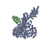
| Title | CryoEM structure of human Mediator subunit Med23 | |||||||||
 Map data Map data | ||||||||||
 Sample Sample |
| |||||||||
 Keywords Keywords | Mediator / MED23 / transcription / human | |||||||||
| Function / homology |  Function and homology information Function and homology informationmediator complex / Generic Transcription Pathway / RSV-host interactions / positive regulation of transcription initiation by RNA polymerase II / RNA polymerase II preinitiation complex assembly / positive regulation of transcription elongation by RNA polymerase II / transcription initiation at RNA polymerase II promoter / PPARA activates gene expression / Transcriptional regulation of white adipocyte differentiation / MLL4 and MLL3 complexes regulate expression of PPARG target genes in adipogenesis and hepatic steatosis ...mediator complex / Generic Transcription Pathway / RSV-host interactions / positive regulation of transcription initiation by RNA polymerase II / RNA polymerase II preinitiation complex assembly / positive regulation of transcription elongation by RNA polymerase II / transcription initiation at RNA polymerase II promoter / PPARA activates gene expression / Transcriptional regulation of white adipocyte differentiation / MLL4 and MLL3 complexes regulate expression of PPARG target genes in adipogenesis and hepatic steatosis / transcription regulator complex / transcription coactivator activity / positive regulation of gene expression / regulation of transcription by RNA polymerase II / regulation of DNA-templated transcription / nucleoplasm / nucleus Similarity search - Function | |||||||||
| Biological species |  Homo sapiens (human) Homo sapiens (human) | |||||||||
| Method | single particle reconstruction / cryo EM / Resolution: 3.1 Å | |||||||||
 Authors Authors | Monte D / Verger A / Lens Z / Villeret V | |||||||||
| Funding support |  France, 1 items France, 1 items
| |||||||||
 Citation Citation | Journal: Nat Commun / Year: 2025 Title: Structural basis of human Mediator recruitment by the phosphorylated transcription factor Elk-1. Authors: Didier Monté / Zoé Lens / Frédérique Dewitte / Marcus Fislage / Marc Aumercier / Alexis Verger / Vincent Villeret /  Abstract: One function of Mediator complex subunit MED23 is to mediate transcriptional activation by the phosphorylated transcription factor Elk-1, in response to the Ras-MAPK signaling pathway. Using ...One function of Mediator complex subunit MED23 is to mediate transcriptional activation by the phosphorylated transcription factor Elk-1, in response to the Ras-MAPK signaling pathway. Using cryogenic electron microscopy, we solve a 3.0 Å structure of human MED23 complexed with the phosphorylated activation domain of Elk-1. Elk-1 binds to MED23 via a hydrophobic sequence PSIHFWSTLSP containing one phosphorylated residue (S383), which forms a tight turn around the central Phenylalanine. Binding of Elk-1 induces allosteric changes in MED23 that propagate to the opposite face of the subunit, resulting in the dynamic behavior of a 19-residue segment, which alters the molecular surface of MED23. We design a specific MED23 mutation (G382F) that disrupts Elk--1 binding and consequently impairs Elk-1-dependent serum-induced activation of target genes in the Ras-Raf-MEK-ERK signaling pathway. The structure provides molecular details and insights into a Mediator subunit-transcription factor interface. | |||||||||
| History |
|
- Structure visualization
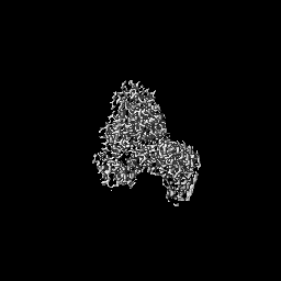
| Supplemental images |
|---|

- Downloads & links
-EMDB archive
| Map data | emd_50247.map.gz | 57.3 MB | EMDB map data format | |
|---|---|---|---|---|
| Header (meta data) | emd-50247-v30.xmlemd-50247.xml | 16.5 KB 16.5 KB | Display Display | EMDB header |
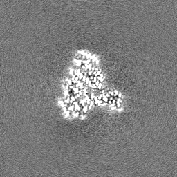
| Images |  emd_50247.png emd_50247.png | 75.2 KB | ||
| Filedesc metadata | emd-50247.cif.gz | 6.5 KB | ||
| Others | emd_50247_half_map_1.map.gzemd_50247_half_map_2.map.gz | 59.4 MB 59.4 MB | ||
| Archive directory |  http://ftp.pdbj.org/pub/emdb/structures/EMD-50247ftp://ftp.pdbj.org/pub/emdb/structures/EMD-50247 http://ftp.pdbj.org/pub/emdb/structures/EMD-50247ftp://ftp.pdbj.org/pub/emdb/structures/EMD-50247 | HTTPS FTP |
-Related structure data
| Related structure data |  9f76MC  9f6yC M: atomic model generated by this map C: citing same article ( |
|---|---|
| Similar structure data |

-Links
| EMDB pages | EMDB (EBI/PDBe) / EMDataResource |
|---|
-Map
| File | Download / File: emd_50247.map.gz / Format: CCP4 / Size: 64 MB / Type: IMAGE STORED AS FLOATING POINT NUMBER (4 BYTES) | ||||||||||||||||||||||||||||||||||||
|---|---|---|---|---|---|---|---|---|---|---|---|---|---|---|---|---|---|---|---|---|---|---|---|---|---|---|---|---|---|---|---|---|---|---|---|---|---|
| Projections & slices | Image control
Images are generated by Spider. | ||||||||||||||||||||||||||||||||||||
| Voxel size | X=Y=Z: 1.01563 Å | ||||||||||||||||||||||||||||||||||||
| Density |
| ||||||||||||||||||||||||||||||||||||
| Symmetry | Space group: 1 | ||||||||||||||||||||||||||||||||||||
| Details | EMDB XML:
|
 Z (Sec.)
 Y (Row.)
 X (Col.)


-Supplemental data
-Half map: #1
| File | emd_50247_half_map_1.map | ||||||||||||
|---|---|---|---|---|---|---|---|---|---|---|---|---|---|
| Projections & Slices |
| ||||||||||||
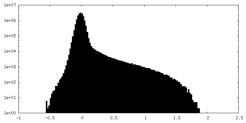
| Density Histograms |


-Half map: #2
| File | emd_50247_half_map_2.map | ||||||||||||
|---|---|---|---|---|---|---|---|---|---|---|---|---|---|
| Projections & Slices |
| ||||||||||||
| Density Histograms |


- Sample components
-Entire : Human Mediator subunit Med23
| Entire | Name: Human Mediator subunit Med23 |
|---|---|
| Components |
|
-Supramolecule #1: Human Mediator subunit Med23
| Supramolecule | Name: Human Mediator subunit Med23 / type: organelle_or_cellular_component / ID: 1 / Parent: 0 / Macromolecule list: all |
|---|---|
| Source (natural) | Organism: Homo sapiens (human) |
-Macromolecule #1: Mediator of RNA polymerase II transcription subunit 23
| Macromolecule | Name: Mediator of RNA polymerase II transcription subunit 23 type: protein_or_peptide / ID: 1 / Details: Med23 sequence with C-terminal His Tag / Number of copies: 1 / Enantiomer: LEVO |
|---|---|
| Source (natural) | Organism: Homo sapiens (human) |
| Molecular weight | Theoretical: 158.294891 KDa |
| Recombinant expression | Organism:   Spodoptera frugiperda (fall armyworm) Spodoptera frugiperda (fall armyworm) |
| Sequence | String: METQLQSIFE EVVKTEVIEE AFPGMFMDTP EDEKTKLISC LGAFRQFWGG LSQESHEQCI QWIVKFIHGQ HSPKRISFLY DCLAMAVET GLLPPRLVCE SLINSDTLEW ERTQLWALTF KLVRKIIGGV DYKGVRDLLK VILEKILTIP NTVSSAVVQQ L LAAREVIA ...String: METQLQSIFE EVVKTEVIEE AFPGMFMDTP EDEKTKLISC LGAFRQFWGG LSQESHEQCI QWIVKFIHGQ HSPKRISFLY DCLAMAVET GLLPPRLVCE SLINSDTLEW ERTQLWALTF KLVRKIIGGV DYKGVRDLLK VILEKILTIP NTVSSAVVQQ L LAAREVIA YILERNACLL PAYFAVTEIR KLYPEGKLPH WLLGNLVSDF VDTFRPTARI NSICGRCSLL PVVNNSGAIC NS WKLDPAT LRFPLKGLLP YDKDLFEPQT ALLRYVLEQP YSRDMVCNML GLNKQHKQRC PVLEDQLVDL VVYAMERSET EEK FDDGGT SQLLWQHLSS QLIFFVLFQF ASFPHMVLSL HQKLAGRGLI KGRDHLMWVL LQFISGSIQK NALADFLPVM KLFD LLYPE KEYIPVPDIN KPQSTHAFAM TCIWIHLNRK AQNDNSKLQI PIPHSLRLHH EFLQQSLRNK SLQMNDYKIA LLCNA YSTN SECFTLPMGA LVETIYGNGI MRIPLPGTNC MASGSITPLP MNLLDSLTVH AKMSLIHSIA TRVIKLAHAK SSVALA PAL VETYSRLLVY MEIESLGIKG FISQLLPTVF KSHAWGILHT LLEMFSYRMH HIQPHYRVQL LSHLHTLAAV AQTNQNQ LH LCVESTALRL ITALGSSEVQ PQFTRFLSDP KTVLSAESEE LNRALILTLA RATHVTDFFT GSDSIQGTWC KDILQTIM S FTPHNWASHT LSCFPGPLQA FFKQNNVPQE SRFNLKKNVE EEYRKWKSMS NENDIITHFS MQGSPPLFLC LLWKMLLET DHINQIGYRV LERIGARALV AHVRTFADFL VYEFSTSAGG QQLNKCIEIL NDMVWKYNIV TLDRLILCLA MRSHEGNEAQ VCYFIIQLL LLKPNDFRNR VSDFVKENSP EHWLQNDWHT KHMNYHKKYP EKLYFEGLAE QVDPPVQIQS PYLPIYFGNV C LRFLPVFD IVIHRFLELL PVSKSLETLL DHLGGLYKFH DRPVTYLYNT LHYYEMHLRD RAFLKRKLVH AIIGSLKDNR PQ GWCLSDT YLKCAMNARE ENPWVPDDTY YCRLIGRLVD TMAGKSPGPF PNCDWRFNEF PNPAAHALHV TCVELMALAV SGK EVGNAL LNVVLKSQPL VPRENITAWM NAIGLIITAL PEPYWIVLHD RIVSVISSPS LTSETEWVGY PFRLFDFTAC HQSY SEMSC SYTLALAHAV WHHSSIGQLS LIPKFLTEVL LPIVKTEFQL LYVYHLVGPF LQRFQQERTR CMIEIGVAFY DMLLN VDQC STHLNYMDPI CDFLYHMKYM FTGDSVKEQV EKIICNLKPA LKLRLRFITH ISKMEPAAVP PQAMNSGSPA PQSNQV PVS LPVTQDVLFQ GPGHHHHHH UniProtKB: Mediator of RNA polymerase II transcription subunit 23 |
-Experimental details
-Structure determination
| Method | cryo EM |
|---|---|
 Processing Processing | single particle reconstruction |
| Aggregation state | particle |
-Sample preparation
| Concentration | 2 mg/mL |
|---|---|
| Buffer | pH: 7.5 / Details: 20mM Tris/HCl, 100mM NaCl, 1mM TCEP |
| Grid | Model: Quantifoil R1.2/1.3 / Material: COPPER / Mesh: 200 / Pretreatment - Type: GLOW DISCHARGE |
| Vitrification | Cryogen name: ETHANE / Chamber humidity: 95 % / Chamber temperature: 277.15 K / Instrument: FEI VITROBOT MARK IV / Details: blotting time 5.6sec, blot force 2. |
- Electron microscopy
| Microscope | TFS KRIOS |
|---|---|
| Image recording | Film or detector model: FEI FALCON IV (4k x 4k) / Average electron dose: 48.0 e/Å2 |
| Electron beam | Acceleration voltage: 300 kV / Electron source:  FIELD EMISSION GUN FIELD EMISSION GUN |
| Electron optics | Illumination mode: FLOOD BEAM / Imaging mode: BRIGHT FIELD / Nominal defocus max: 1.7 µm / Nominal defocus min: 0.8 µm |
| Experimental equipment |  Model: Titan Krios / Image courtesy: FEI Company |