 Movie
Movie Controller


+ Open data

- Basic information
| Entry |  | |||||||||
|---|---|---|---|---|---|---|---|---|---|---|
| Title | Cryo-EM structure of Ch. thermophilum Rai1-Rat1 dimer. | |||||||||
 Map data Map data | ||||||||||
 Sample Sample |
| |||||||||
 Keywords Keywords | Rai1 nuclease / Rtt103 binding / structured / RNA binding / RNA BINDING PROTEIN | |||||||||
| Function / homology |  Function and homology information Function and homology informationmRNA 5'-diphosphatase activity / NAD-cap decapping / 5'-3' RNA exonuclease activity / nuclease activity / nuclear-transcribed mRNA catabolic process / Hydrolases; Acting on acid anhydrides; In phosphorus-containing anhydrides / DNA-templated transcription termination / mRNA processing / rRNA processing / Hydrolases; Acting on ester bonds; Exoribonucleases producing 5'-phosphomonoesters ...mRNA 5'-diphosphatase activity / NAD-cap decapping / 5'-3' RNA exonuclease activity / nuclease activity / nuclear-transcribed mRNA catabolic process / Hydrolases; Acting on acid anhydrides; In phosphorus-containing anhydrides / DNA-templated transcription termination / mRNA processing / rRNA processing / Hydrolases; Acting on ester bonds; Exoribonucleases producing 5'-phosphomonoesters / nucleotide binding / RNA binding / metal ion binding / nucleus / cytosol Similarity search - Function | |||||||||
| Biological species |  Thermochaetoides thermophila (fungus) Thermochaetoides thermophila (fungus) | |||||||||
| Method | single particle reconstruction / cryo EM / Resolution: 3.28 Å | |||||||||
 Authors Authors | Dikunova A / Kubicek K / Noskova N / Stefl R | |||||||||
| Funding support |  Czech Republic, 1 items Czech Republic, 1 items
| |||||||||
 Citation Citation | Journal: Structure / Year: 2025 Title: Assembly of the Xrn2/Rat1-Rai1-Rtt103 termination complexes in mesophilic and thermophilic organisms. Authors: Alzbeta Dikunova / Nikola Noskova / Jan H Overbeck / Martin Polak / David Stelzig / David Zapletal / Karel Kubicek / Jiri Novacek / Remco Sprangers / Richard Stefl /  Abstract: The 5'-3' exoribonuclease Xrn2, known as Rat1 in yeasts, terminates mRNA transcription by RNA polymerase II (RNAPII). In the torpedo model of termination, the activity of Xrn2/Rat1 is enhanced by ...The 5'-3' exoribonuclease Xrn2, known as Rat1 in yeasts, terminates mRNA transcription by RNA polymerase II (RNAPII). In the torpedo model of termination, the activity of Xrn2/Rat1 is enhanced by Rai1, which is recruited to the termination site by Rtt103, an adaptor protein binding to the RNAPII C-terminal domain (CTD). The overall architecture of the Xrn2/Rat1-Rai1-Rtt103 complex remains unknown. We combined structural biology methods to characterize the torpedo complex from Saccharomyces cerevisiae and Chaetomium thermophilum. Comparison of the structures from these organisms revealed a conserved protein core fold of the subunits, but significant variability in their interaction interfaces. We found that in the mesophile, Rtt103 utilizes an unstructured region to augment a Rai1 β-sheet, while in the thermophile Rtt103 binds to a C-terminal helix of Rai1 via its CTD-interacting domain with an α-helical fold. These different torpedo complex assemblies reflect adaptations to the environment and impact complex recruitment to RNAPII. | |||||||||
| History |
|
- Structure visualization
| Supplemental images |
|---|

- Downloads & links
-EMDB archive
| Map data | emd_50048.map.gz | 82.8 MB | EMDB map data format | |
|---|---|---|---|---|
| Header (meta data) | emd-50048-v30.xmlemd-50048.xml | 19.2 KB 19.2 KB | Display Display | EMDB header |
| Images |  emd_50048.png emd_50048.png | 84.6 KB | ||
| Filedesc metadata | emd-50048.cif.gz | 7.1 KB | ||
| Others | emd_50048_half_map_1.map.gzemd_50048_half_map_2.map.gz | 154.5 MB 154.5 MB | ||
| Archive directory |  http://ftp.pdbj.org/pub/emdb/structures/EMD-50048ftp://ftp.pdbj.org/pub/emdb/structures/EMD-50048 http://ftp.pdbj.org/pub/emdb/structures/EMD-50048ftp://ftp.pdbj.org/pub/emdb/structures/EMD-50048 | HTTPS FTP |
-Related structure data
| Related structure data |  9exsMC  8q6vC 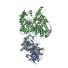 9fmsC C: citing same article ( M: atomic model generated by this map |
|---|---|
| Similar structure data |

-Links
| EMDB pages | EMDB (EBI/PDBe) / EMDataResource |
|---|
-Map
| File | Download / File: emd_50048.map.gz / Format: CCP4 / Size: 166.4 MB / Type: IMAGE STORED AS FLOATING POINT NUMBER (4 BYTES) | ||||||||||||||||||||||||||||||||||||
|---|---|---|---|---|---|---|---|---|---|---|---|---|---|---|---|---|---|---|---|---|---|---|---|---|---|---|---|---|---|---|---|---|---|---|---|---|---|
| Projections & slices | Image control
Images are generated by Spider. | ||||||||||||||||||||||||||||||||||||
| Voxel size | X=Y=Z: 0.834 Å | ||||||||||||||||||||||||||||||||||||
| Density |
| ||||||||||||||||||||||||||||||||||||
| Symmetry | Space group: 1 | ||||||||||||||||||||||||||||||||||||
| Details | EMDB XML:
|
 Z (Sec.)
 Y (Row.)
 X (Col.)


-Supplemental data
-Half map: #2
| File | emd_50048_half_map_1.map | ||||||||||||
|---|---|---|---|---|---|---|---|---|---|---|---|---|---|
| Projections & Slices |
| ||||||||||||
| Density Histograms |


-Half map: #1
| File | emd_50048_half_map_2.map | ||||||||||||
|---|---|---|---|---|---|---|---|---|---|---|---|---|---|
| Projections & Slices |
| ||||||||||||
| Density Histograms |


- Sample components
-Entire : Cryo-EM structure of Ch. thermophilum Rai1-Rat1 complex
| Entire | Name: Cryo-EM structure of Ch. thermophilum Rai1-Rat1 complex |
|---|---|
| Components |
|
-Supramolecule #1: Cryo-EM structure of Ch. thermophilum Rai1-Rat1 complex
| Supramolecule | Name: Cryo-EM structure of Ch. thermophilum Rai1-Rat1 complex type: complex / ID: 1 / Parent: 0 / Macromolecule list: #1-#2 |
|---|---|
| Source (natural) | Organism: Thermochaetoides thermophila (fungus) |
-Macromolecule #1: Decapping nuclease
| Macromolecule | Name: Decapping nuclease / type: protein_or_peptide / ID: 1 / Number of copies: 1 / Enantiomer: LEVO EC number: Hydrolases; Acting on acid anhydrides; In phosphorus-containing anhydrides |
|---|---|
| Source (natural) | Organism: Thermochaetoides thermophila (fungus) |
| Molecular weight | Theoretical: 46.165219 KDa |
| Recombinant expression | Organism:  |
| Sequence | String: MPIEFTIQPP DHYAGVNEPV KRPREFTCFS YDRERRFHLG DRSLKWFYPA YIPSDLSRGY QNWQRHDDSI DEHLDGLLAA IADYEKQTG KPIDAHVTTW RGMMTKIMAT PYDQEEWEMN ATFYRGCIFI EENHAFARRK KMMESSRPAR SDGISPNLMQ Y WGYKFETL ...String: MPIEFTIQPP DHYAGVNEPV KRPREFTCFS YDRERRFHLG DRSLKWFYPA YIPSDLSRGY QNWQRHDDSI DEHLDGLLAA IADYEKQTG KPIDAHVTTW RGMMTKIMAT PYDQEEWEMN ATFYRGCIFI EENHAFARRK KMMESSRPAR SDGISPNLMQ Y WGYKFETL STIPRPWGEV SRDEIESRDD EIVNNMEQYC SVVRTGFGNT IVCLGGEVDA IWDAKPETPG EPINWVELKT SR MITNTGI QTAFDQKLLK YWIQSFLLGV PRIIVGFRDQ DGILRSMEEY ETLNIPYEVR RRGLAKWDGN VCIRFAALFL QWL RLNITE EGVWRIRRPF RGSRIELTKI EQVGHGAIIT EEFMNWRIKL DLQKAKQQAL EEGKQDQGEE GQKAAAPAAA UniProtKB: Decapping nuclease |
-Macromolecule #2: 5'-3' exoribonuclease
| Macromolecule | Name: 5'-3' exoribonuclease / type: protein_or_peptide / ID: 2 / Number of copies: 1 / Enantiomer: LEVO EC number: Hydrolases; Acting on ester bonds; Exoribonucleases producing 5'-phosphomonoesters |
|---|---|
| Source (natural) | Organism: Thermochaetoides thermophila (fungus) |
| Molecular weight | Theoretical: 119.988188 KDa |
| Recombinant expression | Organism: |
| Sequence | String: MGIPAAFRWL SNKYPKIISP VVEERPIVMP DGTEIPVDAT RPNPNGEEFD NLYLDMNGIV HPCSHPEDKP APKDEEEMMI EIFKYTDRI VKMVRPRKIL MIAVDGVAPR AKMNQQRSRR FRAAQEAKEK EEEKKQLLKM LRKEKGSNMQ EEPLETVVKK A FDSNSITP ...String: MGIPAAFRWL SNKYPKIISP VVEERPIVMP DGTEIPVDAT RPNPNGEEFD NLYLDMNGIV HPCSHPEDKP APKDEEEMMI EIFKYTDRI VKMVRPRKIL MIAVDGVAPR AKMNQQRSRR FRAAQEAKEK EEEKKQLLKM LRKEKGSNMQ EEPLETVVKK A FDSNSITP GTPFMDILAA SLRYWCAYKL NTDPAWAKLK VIISDATVPG EGEHKIMEFI RSQRSSPEHN PNTRHVIYGL DA DLIMLGL ATHEPHFRVL REDVFFQEAK ARLCKLCGQK GHDERSCKGE AKQKQGEFDE KDHAQPLKPF IWLHVSILRE YLA AELEVP NLPFRWDLER AIDDWVFLCF FVGNDFLPHL PALEIRENGI DTLTAIWKDN LPIMGGYLTK DGHVDLERAQ YILN GLAKQ EDAIFRRRRE VEERREANAK RRKLNQQGAH AKGAADSHAG KSGRKHVPEA AGPLPGMALF PITNPPPPAI THDMV MKGR SVDQANLANK SAASVLKSQI QSMMAQKAAT NANGAEKDVS ADGTTTAPAS ALGKRKAELI EEDAATNTDT DSVTDG TGS DNEGPVDTVR LWEEGYADRY YEQKFKVDPK DIEFRHKVGR AYAEGLAWVL QYYYQGCPSW EWFYPYHYAP FAADFVD LA KMEIKFEKGR ISRPFEQLMS VLPAASRHAI PEVYHDLMTD PNSPIIDFYP EEFEIDLNGK KMAWQGVALL PFIEMPRL L AAMKEREHLL SEEDRARNEP GFDVLLISDA HPGLYEDITS HFYSKKQGAP KFKLNPRRSD GLAGKVEKIE GYVPHGSLV YPLARNSMPD VDYDRSITVR YIMPSSAHQH KSMLLRGVKL PPPALSRSDI EIIRSKAKNA GRSYGGAPLR NNYNSNGSRR EQPINYAAN APSALPSRNY GSYPGNYGGA NNYGNGYGSG YYPPPGWQPP PPGYPGFGVG VPPPPPPARL AGTPGGYGQG Y GQGYGQGY NQSYGTGYGS GYSSSYQQSA PDRYRPAPAP PPPSTHGYHS GYQSQQSYQG QHHRAGPPLP PSSNNTRRDG RY DDRRGYD DRRDARRDNN PYRDERRYR UniProtKB: 5'-3' exoribonuclease |
-Macromolecule #3: MAGNESIUM ION
| Macromolecule | Name: MAGNESIUM ION / type: ligand / ID: 3 / Number of copies: 1 / Formula: MG |
|---|---|
| Molecular weight | Theoretical: 24.305 Da |
-Experimental details
-Structure determination
| Method | cryo EM |
|---|---|
 Processing Processing | single particle reconstruction |
| Aggregation state | particle |
-Sample preparation
| Concentration | 1.2 mg/mL | ||||||||||||
|---|---|---|---|---|---|---|---|---|---|---|---|---|---|
| Buffer | pH: 7.5 Component:
| ||||||||||||
| Grid | Model: Quantifoil R1.2/1.3 / Material: COPPER / Support film - Material: CARBON / Support film - topology: CONTINUOUS / Pretreatment - Type: GLOW DISCHARGE | ||||||||||||
| Vitrification | Cryogen name: ETHANE / Chamber humidity: 100 % / Chamber temperature: 277.15 K / Instrument: FEI VITROBOT MARK IV |
- Electron microscopy
| Microscope | FEI TITAN KRIOS |
|---|---|
| Image recording | Film or detector model: GATAN K3 (6k x 4k) / Average electron dose: 40.0 e/Å2 |
| Electron beam | Acceleration voltage: 300 kV / Electron source:  FIELD EMISSION GUN FIELD EMISSION GUN |
| Electron optics | Illumination mode: FLOOD BEAM / Imaging mode: BRIGHT FIELD / Nominal defocus max: 3.5 µm / Nominal defocus min: 0.8 µm |
| Experimental equipment |  Model: Titan Krios / Image courtesy: FEI Company |
