 ムービー
ムービー コントローラー


+ データを開く

- 基本情報
基本情報
| 登録情報 |  | ||||||||||||
|---|---|---|---|---|---|---|---|---|---|---|---|---|---|
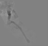
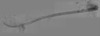
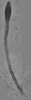
| タイトル | CLEM Cilium N17 for IFT motion study | ||||||||||||
 マップデータ マップデータ | |||||||||||||
 試料 試料 |
| ||||||||||||
 キーワード キーワード | IFT / microtubule / mammalian primary cilia / 3D structure / TRANSPORT PROTEIN | ||||||||||||
| 生物種 |  | ||||||||||||
| 手法 | 電子線トモグラフィー法 / ネガティブ染色法 | ||||||||||||
 データ登録者 データ登録者 | Sun S / Liang B / Koplas A / Tikhonenko I / Nachury M / Khodjakov A / Sui H | ||||||||||||
| 資金援助 |  米国, 3件 米国, 3件
| ||||||||||||
 引用 引用 | ジャーナル: Proc Natl Acad Sci U S A / 年: 2025 タイトル: Intraflagellar transport trains can switch rails and move along multiple microtubules in intact primary cilia. 著者: Shufeng Sun / Biqing Liang / Adam Koplas / Irina Tikhonenko / Maxence Nachury / Alexey Khodjakov / Haixin Sui / 要旨: Structural homeostasis and proper distributions of signaling molecules in cilia require a constant flow of cargoes carried by intraflagellar transport (IFT) trains in both anterograde and retrograde ...Structural homeostasis and proper distributions of signaling molecules in cilia require a constant flow of cargoes carried by intraflagellar transport (IFT) trains in both anterograde and retrograde directions within the thin, long ciliary shafts. In the motile cilium framework, the nine microtubule doublets of the same length serve as the transportation rails, and a preferential association to the two subtubules of the microtubule doublets prevents collisions among the IFT trains that move in opposite directions. However, this mechanism is incompatible with the primary cilia structure, where most of the nine microtubule doublets terminate in the ciliary shafts-only several of them reach the ciliary tip and only in a singlet form. Here, we demonstrate that anterograde and retrograde trains in primary cilia interact with both subtubules of the microtubule doublets without apparent preference. They can switch microtubules, and they may simultaneously interact with multiple microtubules to facilitate their movement. This architecture makes the collisions inevitable, and live-cell recordings reveal that anterograde and retrograde trains tend to pause when they come into direct contact. We also find that the velocity of the train's movement often changes after the pause. Thus, the motion behaviors of IFT trains in primary cilia are distinctive from those of motile cilia, and our data offer an essential foundation for understanding proper signaling molecule distributions in primary cilia. | ||||||||||||
| 履歴 |
|
- 構造の表示
構造の表示
| 添付画像 |
|---|

- ダウンロードとリンク
-EMDBアーカイブ
| マップデータ | emd_49936.map.gz | 4 GB |  EMDBマップデータ形式 EMDBマップデータ形式 | |
|---|---|---|---|---|
| ヘッダ (付随情報) | emd-49936-v30.xmlemd-49936.xml | 14.5 KB 14.5 KB | 表示 表示 | EMDBヘッダ |
| 画像 |  emd_49936.png emd_49936.png | 17.5 KB | ||
| Filedesc metadata | emd-49936.cif.gz | 4.7 KB | ||
| アーカイブディレクトリ |  http://ftp.pdbj.org/pub/emdb/structures/EMD-49936ftp://ftp.pdbj.org/pub/emdb/structures/EMD-49936 http://ftp.pdbj.org/pub/emdb/structures/EMD-49936ftp://ftp.pdbj.org/pub/emdb/structures/EMD-49936 | HTTPS FTP |
-検証レポート
| 文書・要旨 | emd_49936_validation.pdf.gz | 301.9 KB | 表示 | EMDB検証レポート |
|---|---|---|---|---|
| 文書・詳細版 | emd_49936_full_validation.pdf.gz | 301.5 KB | 表示 | |
| XML形式データ | emd_49936_validation.xml.gz | 4.3 KB | 表示 | |
| CIF形式データ | emd_49936_validation.cif.gz | 4.9 KB | 表示 | |
| アーカイブディレクトリ | https://ftp.pdbj.org/pub/emdb/validation_reports/EMD-49936ftp://ftp.pdbj.org/pub/emdb/validation_reports/EMD-49936 | HTTPS FTP |
-関連構造データ

-リンク
| EMDBのページ | EMDB (EBI/PDBe) / EMDataResource |
|---|
-マップ
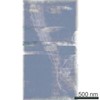
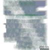
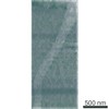
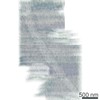
| ファイル | ダウンロード / ファイル: emd_49936.map.gz / 形式: CCP4 / 大きさ: 17.8 GB / タイプ: IMAGE STORED AS SIGNED INTEGER (2 BYTES) | ||||||||||||||||||||||||||||||||
|---|---|---|---|---|---|---|---|---|---|---|---|---|---|---|---|---|---|---|---|---|---|---|---|---|---|---|---|---|---|---|---|---|---|
| 投影像・断面図 | 画像のコントロール
画像は Spider により作成 これらの図は立方格子座標系で作成されたものです | ||||||||||||||||||||||||||||||||
| ボクセルのサイズ | X=Y=Z: 10.624 Å | ||||||||||||||||||||||||||||||||
| 密度 |
| ||||||||||||||||||||||||||||||||
| 対称性 | 空間群: 1 | ||||||||||||||||||||||||||||||||
| 詳細 | EMDB XML:
|
 Z (Sec.)
 Y (Row.)
 X (Col.)


-添付データ
- 試料の構成要素
-全体 : Primary cilium of mIMCD3 cells with triple-mNeonGreen-fusion-IFT88
| 全体 | 名称: Primary cilium of mIMCD3 cells with triple-mNeonGreen-fusion-IFT88 |
|---|---|
| 要素 |
|
-超分子 #1: Primary cilium of mIMCD3 cells with triple-mNeonGreen-fusion-IFT88
| 超分子 | 名称: Primary cilium of mIMCD3 cells with triple-mNeonGreen-fusion-IFT88 タイプ: organelle_or_cellular_component / ID: 1 / 親要素: 0 詳細: This cilium correlates to timed IFT motion tracked by triple-mNeonGreen-fusion-IFT88. This study resulted in kymographic (LM) analysis and subsequent ultrastructure shown here. |
|---|---|
| 由来(天然) | 生物種: |
-実験情報
-構造解析
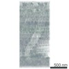
| 手法 | ネガティブ染色法 |
|---|---|
 解析 解析 | 電子線トモグラフィー法 |
| 試料の集合状態 | cell |
-試料調製
| 緩衝液 | pH: 7.4 / 構成要素 - 濃度: 1.0 X / 構成要素 - 式: PBS / 構成要素 - 名称: Phosphate-buffered saline 詳細: NaCl: 137 mM KCl: 2.7 mM Na2HPO4: 10 mM KH2PO4: 1.8 mM |
|---|---|
| 染色 | タイプ: POSITIVE / 材質: Uranyl Acetate, Lead Citrate 詳細: The sections were stained with Uranyl acetate followed by lead citrate solution. |
| 糖包埋 | 材質: SPI-Pon812 詳細: Samples were dehydrated post fixation using an ethanol gradient at RT. Specimens were then back-substituted with acetone. After infiltration with resin the samples were cured. |
| グリッド | モデル: Homemade / 材質: COPPER / 支持フィルム - 材質: FORMVAR / 支持フィルム - トポロジー: CONTINUOUS |
| 詳細 | Specimen is mIMCD3 cells labeled with triple-mNeonGreen-fusion-IFT88. |
| Cryo protectant | no |
| 切片作成 | ウルトラミクロトーム - 装置: UC6 / ウルトラミクロトーム - 温度: 293 K / ウルトラミクロトーム - 最終 厚さ: 120 ウルトラミクロトーム - 詳細: The samples in the resin block were sectioned into serial sections with a target thickness of 150 nm. Most of the sections had an actual thickness of around 120 nm. |
| 位置合わせマーカー | Manufacturer: Sigma / 直径: 10 nm |
- 電子顕微鏡法
| 顕微鏡 | FEI TECNAI F20 |
|---|---|
| 温度 | 最低: 292.0 K / 最高: 298.0 K |
| 詳細 | A Philips High tilt specimen holder was used for data collection purposes. |
| 撮影 | フィルム・検出器のモデル: TVIPS TEMCAM-F416 (4k x 4k) 実像数: 6940 / 平均電子線量: 15.0 e/Å2 |
| 電子線 | 加速電圧: 200 kV / 電子線源:  FIELD EMISSION GUN FIELD EMISSION GUN |
| 電子光学系 | C2レンズ絞り径: 100.0 µm / 最大 デフォーカス(補正後): 4.2 µm / 最小 デフォーカス(補正後): 3.5 µm / 倍率(補正後): 19000 / 照射モード: FLOOD BEAM / 撮影モード: BRIGHT FIELD / Cs: 1.2 mm / 最大 デフォーカス(公称値): 4.0 µm / 最小 デフォーカス(公称値): 3.0 µm / 倍率(公称値): 19000 |
| 試料ステージ | 試料ホルダーモデル: OTHER |
| 実験機器 |  モデル: Tecnai F20 / 画像提供: FEI Company |
-画像解析
| 最終 再構成 | アルゴリズム: SIMULTANEOUS ITERATIVE (SIRT) / ソフトウェア - 名称: TOMO3D / 使用した粒子像数: 6940 |
|---|
