 Movie
Movie Controller


+ Open data

- Basic information
| Entry |  | ||||||||||||
|---|---|---|---|---|---|---|---|---|---|---|---|---|---|
| Title | CLEM Cilium N7 for IFT motion study | ||||||||||||
 Map data Map data | This data was collected as part of a CLEM study of IFT motion (Cilia N7) | ||||||||||||
 Sample Sample |
| ||||||||||||
 Keywords Keywords | IFT / microtubule / mammalian primary cilia / 3D structure / TRANSPORT PROTEIN | ||||||||||||
| Biological species |  | ||||||||||||
| Method | electron tomography / negative staining | ||||||||||||
 Authors Authors | Sun S / Liang B / Koplas A / Tikhonenko I / Nachury M / Khodjakov A / Sui H | ||||||||||||
| Funding support |  United States, 3 items United States, 3 items
| ||||||||||||
 Citation Citation | Journal: Proc Natl Acad Sci U S A / Year: 2025 Title: Intraflagellar transport trains can switch rails and move along multiple microtubules in intact primary cilia. Authors: Shufeng Sun / Biqing Liang / Adam Koplas / Irina Tikhonenko / Maxence Nachury / Alexey Khodjakov / Haixin Sui / Abstract: Structural homeostasis and proper distributions of signaling molecules in cilia require a constant flow of cargoes carried by intraflagellar transport (IFT) trains in both anterograde and retrograde ...Structural homeostasis and proper distributions of signaling molecules in cilia require a constant flow of cargoes carried by intraflagellar transport (IFT) trains in both anterograde and retrograde directions within the thin, long ciliary shafts. In the motile cilium framework, the nine microtubule doublets of the same length serve as the transportation rails, and a preferential association to the two subtubules of the microtubule doublets prevents collisions among the IFT trains that move in opposite directions. However, this mechanism is incompatible with the primary cilia structure, where most of the nine microtubule doublets terminate in the ciliary shafts-only several of them reach the ciliary tip and only in a singlet form. Here, we demonstrate that anterograde and retrograde trains in primary cilia interact with both subtubules of the microtubule doublets without apparent preference. They can switch microtubules, and they may simultaneously interact with multiple microtubules to facilitate their movement. This architecture makes the collisions inevitable, and live-cell recordings reveal that anterograde and retrograde trains tend to pause when they come into direct contact. We also find that the velocity of the train's movement often changes after the pause. Thus, the motion behaviors of IFT trains in primary cilia are distinctive from those of motile cilia, and our data offer an essential foundation for understanding proper signaling molecule distributions in primary cilia. | ||||||||||||
| History |
|
- Structure visualization
| Supplemental images |
|---|

- Downloads & links
-EMDB archive
| Map data | emd_49937.map.gz | 7.6 GB |  EMDB map data format EMDB map data format | |
|---|---|---|---|---|
| Header (meta data) | emd-49937-v30.xmlemd-49937.xml | 14.6 KB 14.6 KB | Display Display | EMDB header |
| Images |  emd_49937.png emd_49937.png | 18.4 KB | ||
| Filedesc metadata | emd-49937.cif.gz | 4.6 KB | ||
| Archive directory |  http://ftp.pdbj.org/pub/emdb/structures/EMD-49937ftp://ftp.pdbj.org/pub/emdb/structures/EMD-49937 http://ftp.pdbj.org/pub/emdb/structures/EMD-49937ftp://ftp.pdbj.org/pub/emdb/structures/EMD-49937 | HTTPS FTP |
-Related structure data
| Related structure data | C: citing same article ( |
|---|
-Links
| EMDB pages | EMDB (EBI/PDBe) / EMDataResource |
|---|
-Map
| File | Download / File: emd_49937.map.gz / Format: CCP4 / Size: 47 GB / Type: IMAGE STORED AS SIGNED INTEGER (2 BYTES) | ||||||||||||||||||||||||||||||||
|---|---|---|---|---|---|---|---|---|---|---|---|---|---|---|---|---|---|---|---|---|---|---|---|---|---|---|---|---|---|---|---|---|---|
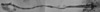
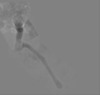
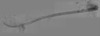
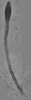
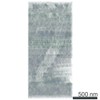
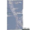
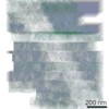
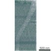
| Annotation | This data was collected as part of a CLEM study of IFT motion (Cilia N7) | ||||||||||||||||||||||||||||||||
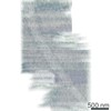
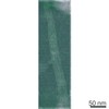
| Projections & slices | Image control
Images are generated by Spider. generated in cubic-lattice coordinate | ||||||||||||||||||||||||||||||||
| Voxel size | X=Y=Z: 10.25 Å | ||||||||||||||||||||||||||||||||
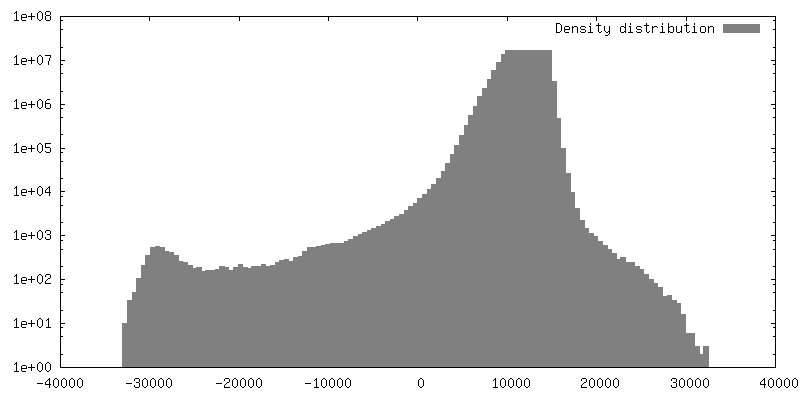
| Density |
| ||||||||||||||||||||||||||||||||
| Symmetry | Space group: 1 | ||||||||||||||||||||||||||||||||
| Details | EMDB XML:
|
 Z (Sec.)
 Y (Row.)
 X (Col.)


-Supplemental data
- Sample components
-Entire : Primary cilium of mIMCD3 cells with triple-mNeonGreen-fusion-IFT88
| Entire | Name: Primary cilium of mIMCD3 cells with triple-mNeonGreen-fusion-IFT88 |
|---|---|
| Components |
|
-Supramolecule #1: Primary cilium of mIMCD3 cells with triple-mNeonGreen-fusion-IFT88
| Supramolecule | Name: Primary cilium of mIMCD3 cells with triple-mNeonGreen-fusion-IFT88 type: organelle_or_cellular_component / ID: 1 / Parent: 0 Details: This cilium correlates to timed IFT motion tracked by triple-mNeonGreen-fusion-IFT88. This study resulted in kymographic (LM) analysis and subsequent ultrastructure shown here. |
|---|---|
| Source (natural) | Organism: |
-Experimental details
-Structure determination
| Method | negative staining |
|---|---|
 Processing Processing | electron tomography |
| Aggregation state | cell |
-Sample preparation
| Buffer | pH: 7.4 / Component - Concentration: 1.0 X / Component - Formula: PBS / Component - Name: Phosphate-buffered saline Details: NaCl: 137 mM KCl: 2.7 mM Na2HPO4: 10 mM KH2PO4: 1.8 mM |
|---|---|
| Staining | Type: POSITIVE / Material: Uranyl Acetate/Lead Citrate Details: The sections were stained with Uranyl acetate followed by lead citrate solution. |
| Sugar embedding | Material: SPI-Pon812 Details: Samples were dehydrated post fixation using an ethanol gradient at RT. Specimens are then back-substituted with acetone. After infiltration with resin the samples were cured. |
| Grid | Model: Homemade / Material: COPPER / Support film - Material: FORMVAR / Support film - topology: CONTINUOUS |
| Details | Specimen is mIMCD3 cells labeled with triple-mNeonGreen-fusion-IFT88. |
| Sectioning | Ultramicrotomy - Instrument: UC6 / Ultramicrotomy - Temperature: 293 K / Ultramicrotomy - Final thickness: 120 Ultramicrotomy - Details: The samples in the resin block were sectioned into serial sections with a target thickness of 150nm. Most sections had an actual thickness of around 120nm. |
| Fiducial marker | Manufacturer: sigma / Diameter: 10 nm |
- Electron microscopy
| Microscope | FEI TECNAI F20 |
|---|---|
| Temperature | Min: 292.0 K / Max: 298.0 K |
| Image recording | Film or detector model: TVIPS TEMCAM-F416 (4k x 4k) / Average electron dose: 15.0 e/Å2 |
| Electron beam | Acceleration voltage: 200 kV / Electron source:  FIELD EMISSION GUN FIELD EMISSION GUN |
| Electron optics | C2 aperture diameter: 100.0 µm / Calibrated defocus max: 3.5 µm / Calibrated defocus min: 2.5 µm / Calibrated magnification: 19000 / Illumination mode: FLOOD BEAM / Imaging mode: BRIGHT FIELD / Cs: 1.2 mm / Nominal defocus max: 4.0 µm / Nominal defocus min: 3.0 µm / Nominal magnification: 19000 |
| Sample stage | Specimen holder model: OTHER |
| Experimental equipment |  Model: Tecnai F20 / Image courtesy: FEI Company |
-Image processing
| Final reconstruction | Algorithm: SIMULTANEOUS ITERATIVE (SIRT) / Software - Name: TOMO3D / Number images used: 13400 |
|---|
