Movie
Movie Controller

[English] 日本語
 Yorodumi
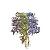
- EMDB-49840: ATPase Hybrid F1 with the ancestral core domains Catalytic Dwell -
+ Open data

- Basic information
| Entry |  | |||||||||
|---|---|---|---|---|---|---|---|---|---|---|
| Title | ATPase Hybrid F1 with the ancestral core domains Catalytic Dwell | |||||||||
 Map data Map data | ATPase Hybrid F1 with the ancestral core domains Catalytic Dwell | |||||||||
 Sample Sample |
| |||||||||
 Keywords Keywords | Enerygy / Ancestral ATPase / ELECTRON TRANSPORT | |||||||||
| Function / homology |  Function and homology information Function and homology informationproton motive force-driven plasma membrane ATP synthesis / proton-transporting ATP synthase complex / proton-transporting ATP synthase activity, rotational mechanism / ATP binding / plasma membrane Similarity search - Function | |||||||||
| Biological species |   Pseudomonas aeruginosa (bacteria) Pseudomonas aeruginosa (bacteria) | |||||||||
| Method | single particle reconstruction / cryo EM / Resolution: 2.58 Å | |||||||||
 Authors Authors | Stewart AG / Noji H / Sobti M / Suzuki AK | |||||||||
| Funding support |  Australia, 1 items Australia, 1 items
| |||||||||
 Citation Citation | Journal: Protein Sci / Year: 2025 Title: Functional and structural characterization of F-ATPase with common ancestral core domains in stator ring. Authors: Aya K Suzuki / Ryutaro Furukawa / Meghna Sobti / Simon H J Brown / Alastair G Stewart / Satoshi Akanuma / Hiroshi Ueno / Hiroyuki Noji /  Abstract: Extant F-ATPases exhibit diverse rotational stepping behaviors-3-, 6-, or 9-step cycles-yet the evolutionary origin of these patterns remains unclear. Here, we used ancestral sequence reconstruction ...Extant F-ATPases exhibit diverse rotational stepping behaviors-3-, 6-, or 9-step cycles-yet the evolutionary origin of these patterns remains unclear. Here, we used ancestral sequence reconstruction to infer the catalytic β and non-catalytic α subunits of a putative ancestral F-ATPase. We then fused their functionally critical domains into the thermostable F from Bacillus PS3, yielding a stable chimeric enzyme. Cryo-EM revealed two distinct conformational states-binding and catalytic dwell states-separated by a ~34° rotation of the γ subunit, suggesting a fundamental six-step mechanism akin to that of extant six-stepping F-ATPases. Single-molecule rotation assays with ATP and the slowly hydrolyzed ATP analog ATPγS demonstrated that the chimeric motor is intrinsically a six-stepper, pausing at binding and catalytic dwell positions separated by 32.1°, although the binding dwell is significantly prolonged by an unknown mechanism. These findings indicate that F-ATPase was originally a six-stepper and diversified into 3-, 6- and 9-step forms in evolutionary adaptation. Based on these results, we discuss plausible features of the entire FF complex, along with potential physiological contexts in the last universal common ancestor and related lineages. | |||||||||
| History |
|
- Structure visualization
| Supplemental images |
|---|

- Downloads & links
-EMDB archive
| Map data | emd_49840.map.gz | 59.6 MB | EMDB map data format | |
|---|---|---|---|---|
| Header (meta data) | emd-49840-v30.xmlemd-49840.xml | 20.8 KB 20.8 KB | Display Display | EMDB header |
| FSC (resolution estimation) | emd_49840_fsc.xml | 8.4 KB | Display | FSC data file |
| Images |  emd_49840.png emd_49840.png | 65.7 KB | ||
| Filedesc metadata | emd-49840.cif.gz | 6.4 KB | ||
| Others | emd_49840_additional_1.map.gzemd_49840_half_map_1.map.gzemd_49840_half_map_2.map.gz | 53.8 MB 59.4 MB 59.4 MB | ||
| Archive directory |  http://ftp.pdbj.org/pub/emdb/structures/EMD-49840ftp://ftp.pdbj.org/pub/emdb/structures/EMD-49840 http://ftp.pdbj.org/pub/emdb/structures/EMD-49840ftp://ftp.pdbj.org/pub/emdb/structures/EMD-49840 | HTTPS FTP |
-Related structure data
| Related structure data |  9nvmMC  9nvlC M: atomic model generated by this map C: citing same article ( |
|---|---|
| Similar structure data |


-Links
| EMDB pages | EMDB (EBI/PDBe) / EMDataResource |
|---|---|
| Related items in Molecule of the Month |


-Map
| File | Download / File: emd_49840.map.gz / Format: CCP4 / Size: 64 MB / Type: IMAGE STORED AS FLOATING POINT NUMBER (4 BYTES) | ||||||||||||||||||||||||||||||||||||
|---|---|---|---|---|---|---|---|---|---|---|---|---|---|---|---|---|---|---|---|---|---|---|---|---|---|---|---|---|---|---|---|---|---|---|---|---|---|
| Annotation | ATPase Hybrid F1 with the ancestral core domains Catalytic Dwell | ||||||||||||||||||||||||||||||||||||
| Projections & slices | Image control
Images are generated by Spider. | ||||||||||||||||||||||||||||||||||||
| Voxel size | X=Y=Z: 0.84 Å | ||||||||||||||||||||||||||||||||||||
| Density |
| ||||||||||||||||||||||||||||||||||||
| Symmetry | Space group: 1 | ||||||||||||||||||||||||||||||||||||
| Details | EMDB XML:
|
 Z (Sec.)
 Y (Row.)
 X (Col.)


-Supplemental data
-Additional map: Additional Map
| File | emd_49840_additional_1.map | ||||||||||||
|---|---|---|---|---|---|---|---|---|---|---|---|---|---|
| Annotation | Additional Map | ||||||||||||
| Projections & Slices |
| ||||||||||||
| Density Histograms |


-Half map: Half Map B
| File | emd_49840_half_map_1.map | ||||||||||||
|---|---|---|---|---|---|---|---|---|---|---|---|---|---|
| Annotation | Half Map B | ||||||||||||
| Projections & Slices |
| ||||||||||||
| Density Histograms |


-Half map: Half Map A
| File | emd_49840_half_map_2.map | ||||||||||||
|---|---|---|---|---|---|---|---|---|---|---|---|---|---|
| Annotation | Half Map A | ||||||||||||
| Projections & Slices |
| ||||||||||||
| Density Histograms |


- Sample components
-Entire : ATPase Hybrid F1 with the ancestral core domains
| Entire | Name: ATPase Hybrid F1 with the ancestral core domains |
|---|---|
| Components |
|
-Supramolecule #1: ATPase Hybrid F1 with the ancestral core domains
| Supramolecule | Name: ATPase Hybrid F1 with the ancestral core domains / type: complex / ID: 1 / Parent: 0 / Macromolecule list: #1-#3 |
|---|---|
| Source (natural) | Organism: |
-Macromolecule #1: ATPase hybrid F1 with the ancestral core domains alpha chains
| Macromolecule | Name: ATPase hybrid F1 with the ancestral core domains alpha chains type: protein_or_peptide / ID: 1 / Number of copies: 3 / Enantiomer: LEVO |
|---|---|
| Source (natural) | Organism: |
| Molecular weight | Theoretical: 55.987812 KDa |
| Recombinant expression | Organism: |
| Sequence | String: MSHHHHHHGS IRAEEISALI KQQIENYESQ IQVSDVGTVI QVGDGIARAH GLDNVMSGEL VEFANGVMGM ALNLEENNVG IVILGPYTG IKEGDEVRRT GRIMEVPVGE ELIGRVVNAL GQPIDGKGPI NAKEFRPVER KAPGVVDRQP VKEPLQTGIK A IDAMIPIG ...String: MSHHHHHHGS IRAEEISALI KQQIENYESQ IQVSDVGTVI QVGDGIARAH GLDNVMSGEL VEFANGVMGM ALNLEENNVG IVILGPYTG IKEGDEVRRT GRIMEVPVGE ELIGRVVNAL GQPIDGKGPI NAKEFRPVER KAPGVVDRQP VKEPLQTGIK A IDAMIPIG RGQRELIIGD RQTGKTAIAI DTIINQKGQD VICIYVAIGQ KQSTVAQVVK TLEEHGAMEY TIVVAATASD PA ALQYIAP YAGCAMGEYF RDKGKHALVV YDDLSKHAVA YRQISLLLRR PPGREAYPGD VFYLHSRLLE RAAKLSDEKG GGS LTALPI IETQAGDVSA YIPTNVISIT DGQIYLESDL FYSGIRPAIN VGLSVSRVGG AAQIKAMKQV AGKLRLDLAQ YREL QAFAQ FASDLDEATR AQLERGQRMT EILKQPQYSP MPVEKQVVII YAGTNGYLDD IPVEKVKEFE DGFLEYIESK HPDIL EEIR EKKALDDELE EKLKKAIKEF KATFK |
-Macromolecule #2: ATPase hybrid F1 with the ancestral core domains beta chains
| Macromolecule | Name: ATPase hybrid F1 with the ancestral core domains beta chains type: protein_or_peptide / ID: 2 / Number of copies: 3 / Enantiomer: LEVO |
|---|---|
| Source (natural) | Organism: |
| Molecular weight | Theoretical: 53.888594 KDa |
| Recombinant expression | Organism: |
| Sequence | String: MHHHHHHHHH HMTRGRVIQV MGPVVDVKFE NGHLPAIYNA LKIQHKARNE NEVDIDLTLE VALHLGDDTV RTIAMASTDG LIRGMEVID TGAPISVPVG PETLGRMFDV LGEPIDEKGP VKAKKRWPIH RPPPSLSEQS TEDEILETGI KVIDLLAPIP K GGKIGLFG ...String: MHHHHHHHHH HMTRGRVIQV MGPVVDVKFE NGHLPAIYNA LKIQHKARNE NEVDIDLTLE VALHLGDDTV RTIAMASTDG LIRGMEVID TGAPISVPVG PETLGRMFDV LGEPIDEKGP VKAKKRWPIH RPPPSLSEQS TEDEILETGI KVIDLLAPIP K GGKIGLFG GAGVGKTVLI MELIRNIAYE HKGFSVFAGV GERSREGNEL WLEMKESGVL DNTVLVFGQM NEPPGARFRV AL TGLTMAE YFRDEEGKDV LLFIDNIFRF AQAGSEVSAL LGRMPSEVGY QPTLATEMAE LQERITSTRR GSITSVQAIY VPA DDLTDP APATTFAHLD ATIVLSRELA EKGIYPAVDP LQSTSRIMDP RIVSEEHYEV ARRVREILQR YKDLQDIIAI LGME ELSEE DKLIVQRARK IQRFLSQPFH VAEHFTGRPG KYVPIEDTIR GFKEILDGKL DDVPEQAFYM VGTIEEAVEK AKKMK KE |
-Macromolecule #3: ATP synthase gamma chain
| Macromolecule | Name: ATP synthase gamma chain / type: protein_or_peptide / ID: 3 / Number of copies: 1 / Enantiomer: LEVO |
|---|---|
| Source (natural) | Organism: Pseudomonas aeruginosa (bacteria) |
| Molecular weight | Theoretical: 31.86557 KDa |
| Recombinant expression | Organism: |
| Sequence | String: MASLRDIKTR INATKKTSQI TKAMEMVSTS KLNRAEQNAK SFVPYMEKIQ EVVANVALGA GGASHPMLVS RPVKKTGYLV ITSDRGLAG AYNSNVLRLV YQTIQKRHAC PDEYAIIVIG RVGLSFFRKR NMPVILDITR LPDQPSFADI KEIARKTVGL F ADGTFDEL ...String: MASLRDIKTR INATKKTSQI TKAMEMVSTS KLNRAEQNAK SFVPYMEKIQ EVVANVALGA GGASHPMLVS RPVKKTGYLV ITSDRGLAG AYNSNVLRLV YQTIQKRHAC PDEYAIIVIG RVGLSFFRKR NMPVILDITR LPDQPSFADI KEIARKTVGL F ADGTFDEL YMYYNHYVSA IQQEVTERKL LPLTDLAENK QRTVYEFEPS QEECLDVLLP QYAESLIYGA LLDAKASEHA AR MTAMKNA TDNANELIRT LTLSYNRARQ AAITQEITEI VAGANALQ UniProtKB: ATP synthase gamma chain |
-Macromolecule #4: ADENOSINE-5'-TRIPHOSPHATE
| Macromolecule | Name: ADENOSINE-5'-TRIPHOSPHATE / type: ligand / ID: 4 / Number of copies: 3 / Formula: ATP |
|---|---|
| Molecular weight | Theoretical: 507.181 Da |
| Chemical component information |  ChemComp-ATP: |
-Macromolecule #5: MAGNESIUM ION
| Macromolecule | Name: MAGNESIUM ION / type: ligand / ID: 5 / Number of copies: 4 / Formula: MG |
|---|---|
| Molecular weight | Theoretical: 24.305 Da |
-Macromolecule #6: ADENOSINE-5'-DIPHOSPHATE
| Macromolecule | Name: ADENOSINE-5'-DIPHOSPHATE / type: ligand / ID: 6 / Number of copies: 1 / Formula: ADP |
|---|---|
| Molecular weight | Theoretical: 427.201 Da |
| Chemical component information |  ChemComp-ADP: |
-Experimental details
-Structure determination
| Method | cryo EM |
|---|---|
 Processing Processing | single particle reconstruction |
| Aggregation state | particle |
-Sample preparation
| Buffer | pH: 7 |
|---|---|
| Vitrification | Cryogen name: ETHANE |
- Electron microscopy
| Microscope | TFS KRIOS |
|---|---|
| Image recording | Film or detector model: GATAN K3 (6k x 4k) / Average electron dose: 62.0 e/Å2 |
| Electron beam | Acceleration voltage: 300 kV / Electron source:  FIELD EMISSION GUN FIELD EMISSION GUN |
| Electron optics | Illumination mode: FLOOD BEAM / Imaging mode: BRIGHT FIELD / Nominal defocus max: 1.0 µm / Nominal defocus min: 0.2 µm |
| Experimental equipment |  Model: Titan Krios / Image courtesy: FEI Company |