- EMDB-4919: STRUCTURE OF THE MECHANOSENSITIVE CHANNEL MSCS EMBEDDED IN THE ME... -
+
Open data
ID or keywords:
Loading...
-
Basic information
Entry
Database: EMDB / ID: EMD-4919
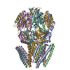
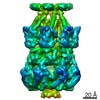
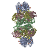
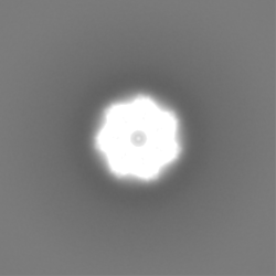
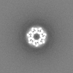
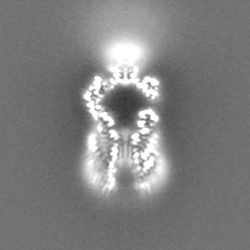
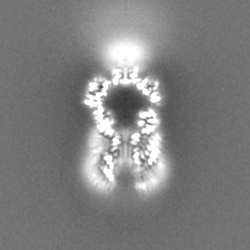
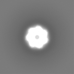
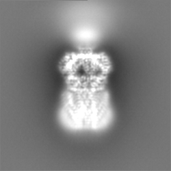
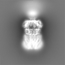
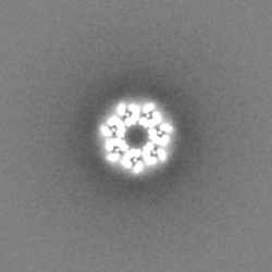
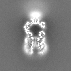
Title
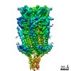
STRUCTURE OF THE MECHANOSENSITIVE CHANNEL MSCS EMBEDDED IN THE MEMBRANE BILAYER
Map data
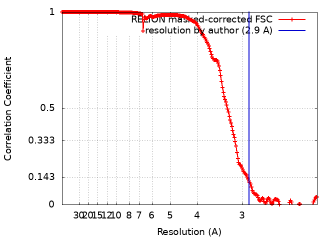
RELION post-processed map; no mask; applied b-factor of -139; new box 250 px
Sample
Complex: MSCS IN NANODISCS
Protein or peptide: Small-conductance mechanosensitive channel
Ligand: 1,2-DIOLEOYL-SN-GLYCERO-3-PHOSPHOCHOLINE
Keywords
MEMBRANE PROTEIN / CHANNEL / MECHANOSENSITIVE
Function / homology
Function and homology information
intracellular water homeostasis / mechanosensitive monoatomic ion channel activity / protein homooligomerization / monoatomic ion transmembrane transport / membrane / identical protein binding / plasma membrane Similarity search - Function
Conserved TM helix / Mechanosensitive ion channel, conserved TM helix / Mechanosensitive ion channel MscS, archaea/bacteria type / : / : / Mechanosensitive ion channel MscS, C-terminal / Mechanosensitive ion channel, transmembrane helices 2/3 / Mechanosensitive ion channel MscS, conserved site / Uncharacterized protein family UPF0003 signature. / Mechanosensitive ion channel MscS, C-terminal ...Conserved TM helix / Mechanosensitive ion channel, conserved TM helix / Mechanosensitive ion channel MscS, archaea/bacteria type / : / : / Mechanosensitive ion channel MscS, C-terminal / Mechanosensitive ion channel, transmembrane helices 2/3 / Mechanosensitive ion channel MscS, conserved site / Uncharacterized protein family UPF0003 signature. / Mechanosensitive ion channel MscS, C-terminal / Mechanosensitive ion channel MscS, transmembrane-2 / Mechanosensitive ion channel MscS / Mechanosensitive ion channel, beta-domain / Mechanosensitive ion channel MscS, beta-domain superfamily / LSM domain superfamily Similarity search - Domain/homology
Journal: J Mol Biol / Year: 2019 Title: Structure of the Mechanosensitive Channel MscS Embedded in the Membrane Bilayer. Authors: Tim Rasmussen / Vanessa J Flegler / Akiko Rasmussen / Bettina Böttcher / Abstract: Since life has emerged, gradients of osmolytes over the cell membrane cause pressure changes in the cell and require tight regulation to prevent cell rupture. The mechanosensitive channel of small ...Since life has emerged, gradients of osmolytes over the cell membrane cause pressure changes in the cell and require tight regulation to prevent cell rupture. The mechanosensitive channel of small conductance (MscS) releases solutes and water when a hypo-osmotic shock raises the pressure in the cell. It is a member of a large family of MscS-like channels found in bacteria, archaea, fungi and plants and model for mechanosensation. MscS senses the increase of tension in the membrane directly by the force from the lipids, but the molecular mechanism is still elusive. We determined the lipid interactions of MscS by resolving the structure of Escherichia coli MscS embedded in membrane discs to 2.9-Å resolution using cryo-electron microscopy. The membrane is attached only to parts of the sensor paddles of MscS, but phospholipid molecules move through grooves into remote pockets on the cytosolic side. On the periplasmic side, a lipid bound by R88 at the pore entrance is separated from the membrane by TM1 helices. The N-terminus interacts with the periplasmic membrane surface. We demonstrate that the unique membrane domain of MscS promotes deep penetration of lipid molecules and shows multimodal interaction with the membrane to fine-tune tension sensing.
History
Deposition
May 2, 2019
-
Header (metadata) release
Jun 12, 2019
-
Map release
Jul 24, 2019
-
Update
May 22, 2024
-
Current status
May 22, 2024
Processing site: PDBe / Status: Released
-
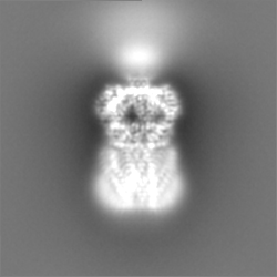
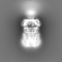
Structure visualization
Movie
Surface view with section colored by density value
In the structure databanks used in Yorodumi, some data are registered as the other names, "COVID-19 virus" and "2019-nCoV". Here are the details of the virus and the list of structure data.
Jan 31, 2019. EMDB accession codes are about to change! (news from PDBe EMDB page)
EMDB accession codes are about to change! (news from PDBe EMDB page)
The allocation of 4 digits for EMDB accession codes will soon come to an end. Whilst these codes will remain in use, new EMDB accession codes will include an additional digit and will expand incrementally as the available range of codes is exhausted. The current 4-digit format prefixed with “EMD-” (i.e. EMD-XXXX) will advance to a 5-digit format (i.e. EMD-XXXXX), and so on. It is currently estimated that the 4-digit codes will be depleted around Spring 2019, at which point the 5-digit format will come into force.
The EM Navigator/Yorodumi systems omit the EMD- prefix.
Related info.:Q: What is EMD? / ID/Accession-code notation in Yorodumi/EM Navigator
Yorodumi is a browser for structure data from EMDB, PDB, SASBDB, etc.
This page is also the successor to EM Navigator detail page, and also detail information page/front-end page for Omokage search.
The word "yorodu" (or yorozu) is an old Japanese word meaning "ten thousand". "mi" (miru) is to see.
Related info.:EMDB / PDB / SASBDB / Comparison of 3 databanks / Yorodumi Search / Aug 31, 2016. New EM Navigator & Yorodumi / Yorodumi Papers / Jmol/JSmol / Function and homology information / Changes in new EM Navigator and Yorodumi
 Movie
Movie Controller

 Yorodumi
 Open data
 Basic information
 Map data
 Sample
 Keywords
 Function and homology information
 Authors
 Germany, 2 items
  Citation
 Structure visualization
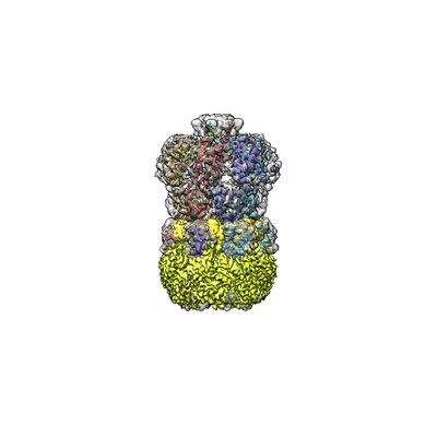
 Downloads & links
 emd_4919.png
 http://ftp.pdbj.org/pub/emdb/structures/EMD-4919
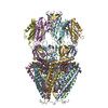


 Z (Sec.)
 Y (Row.)
 X (Col.)


 Sample components
 Processing
 Electron microscopy
 FIELD EMISSION GUN