Movie
Movie Controller

[English] 日本語
 Yorodumi
- PDB-6rld: STRUCTURE OF THE MECHANOSENSITIVE CHANNEL MSCS EMBEDDED IN THE ME... -
+ Open data

- Basic information
| Entry | Database: PDB / ID: 6rld | |||||||||
|---|---|---|---|---|---|---|---|---|---|---|
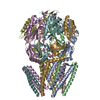
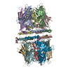
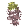
| Title | STRUCTURE OF THE MECHANOSENSITIVE CHANNEL MSCS EMBEDDED IN THE MEMBRANE BILAYER | |||||||||
 Components Components | Small-conductance mechanosensitive channel | |||||||||
 Keywords Keywords | MEMBRANE PROTEIN / CHANNEL / MECHANOSENSITIVE | |||||||||
| Function / homology |  Function and homology information Function and homology informationintracellular water homeostasis / mechanosensitive monoatomic ion channel activity / protein homooligomerization / monoatomic ion transmembrane transport / membrane / identical protein binding / plasma membrane Similarity search - Function | |||||||||
| Biological species |  | |||||||||
| Method | ELECTRON MICROSCOPY / single particle reconstruction / Resolution: 2.9 Å | |||||||||
 Authors Authors | Rasmussen, T. / Flegler, V.J. / Rasmussen, A. / Boettcher, B. | |||||||||
| Funding support |  Germany, 2items Germany, 2items
| |||||||||
 Citation Citation | Journal: J Mol Biol / Year: 2019 Title: Structure of the Mechanosensitive Channel MscS Embedded in the Membrane Bilayer. Authors: Tim Rasmussen / Vanessa J Flegler / Akiko Rasmussen / Bettina Böttcher / Abstract: Since life has emerged, gradients of osmolytes over the cell membrane cause pressure changes in the cell and require tight regulation to prevent cell rupture. The mechanosensitive channel of small ...Since life has emerged, gradients of osmolytes over the cell membrane cause pressure changes in the cell and require tight regulation to prevent cell rupture. The mechanosensitive channel of small conductance (MscS) releases solutes and water when a hypo-osmotic shock raises the pressure in the cell. It is a member of a large family of MscS-like channels found in bacteria, archaea, fungi and plants and model for mechanosensation. MscS senses the increase of tension in the membrane directly by the force from the lipids, but the molecular mechanism is still elusive. We determined the lipid interactions of MscS by resolving the structure of Escherichia coli MscS embedded in membrane discs to 2.9-Å resolution using cryo-electron microscopy. The membrane is attached only to parts of the sensor paddles of MscS, but phospholipid molecules move through grooves into remote pockets on the cytosolic side. On the periplasmic side, a lipid bound by R88 at the pore entrance is separated from the membrane by TM1 helices. The N-terminus interacts with the periplasmic membrane surface. We demonstrate that the unique membrane domain of MscS promotes deep penetration of lipid molecules and shows multimodal interaction with the membrane to fine-tune tension sensing. | |||||||||
| History |
|
- Structure visualization
| Movie |
Movie viewer |
|---|---|
| Structure viewer | Molecule: MolmilJmol/JSmol |
- Downloads & links
-Download
| PDBx/mmCIF format | 6rld.cif.gz | 331.8 KB | Display | PDBx/mmCIF format |
|---|---|---|---|---|
| PDB format | pdb6rld.ent.gz | 280.3 KB | Display | PDB format |
| PDBx/mmJSON format | 6rld.json.gz | Tree view | PDBx/mmJSON format | |
| Others |  Other downloads Other downloads |
-Validation report
| Arichive directory | https://data.pdbj.org/pub/pdb/validation_reports/rl/6rldftp://data.pdbj.org/pub/pdb/validation_reports/rl/6rld | HTTPS FTP |
|---|
-Related structure data
| Related structure data | 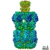 4919MC M: map data used to model this data C: citing same article ( |
|---|---|
| Similar structure data |


-Links
 PDBj
PDBj

- Assembly
| Deposited unit | 
|
|---|---|
| 1 |
|
-Components
| #1: Protein | Mass: 30922.898 Da / Num. of mol.: 7 / Source method: isolated from a natural source / Source: (natural) #2: Chemical | ChemComp-PCW /   Mass: 787.121 Da / Num. of mol.: 21 / Source method: obtained synthetically / Formula: C44H85NO8P / Feature type: SUBJECT OF INVESTIGATION / Comment: DOPC, phospholipid*YM Mass: 787.121 Da / Num. of mol.: 21 / Source method: obtained synthetically / Formula: C44H85NO8P / Feature type: SUBJECT OF INVESTIGATION / Comment: DOPC, phospholipid*YM |
|---|
-Experimental details
-Experiment
| Experiment | Method: ELECTRON MICROSCOPY |
|---|---|
| EM experiment | Aggregation state: PARTICLE / 3D reconstruction method: single particle reconstruction |
- Sample preparation
| Component | Name: MSCS IN NANODISCS / Type: COMPLEX / Details: MSCS WITH MSP1E3D1 AND AZOLECTINE / Entity ID: #1 / Source: NATURAL |
|---|---|
| Source (natural) | Organism: |
| Buffer solution | pH: 7.5 |
| Specimen | Conc.: 0.4 mg/ml / Embedding applied: NO / Shadowing applied: NO / Staining applied: NO / Vitrification applied: NO |
| Specimen support | Details: QUANTIFOIL R1.2/1.3 |
- Electron microscopy imaging
| Experimental equipment |  Model: Titan Krios / Image courtesy: FEI Company | ||||||||||||||||||||||||||||||
|---|---|---|---|---|---|---|---|---|---|---|---|---|---|---|---|---|---|---|---|---|---|---|---|---|---|---|---|---|---|---|---|
| Microscopy | Model: FEI TITAN KRIOS | ||||||||||||||||||||||||||||||
| Electron gun | Electron source:  FIELD EMISSION GUN / Accelerating voltage: 300 kV / Illumination mode: FLOOD BEAM FIELD EMISSION GUN / Accelerating voltage: 300 kV / Illumination mode: FLOOD BEAM | ||||||||||||||||||||||||||||||
| Electron lens | Mode: BRIGHT FIELD / Nominal magnification: 75000 X / Nominal defocus max: 2400 nm / Nominal defocus min: 900 nm / Cs: 2.7 mm / C2 aperture diameter: 70 µm / Alignment procedure: COMA FREE | ||||||||||||||||||||||||||||||
| Specimen holder | Cryogen: NITROGEN / Specimen holder model: FEI TITAN KRIOS AUTOGRID HOLDER | ||||||||||||||||||||||||||||||
| Image recording |
|
- Processing
| EM software |
| ||||||||||||||||||||||||||||||||||||||||||||||||||
|---|---|---|---|---|---|---|---|---|---|---|---|---|---|---|---|---|---|---|---|---|---|---|---|---|---|---|---|---|---|---|---|---|---|---|---|---|---|---|---|---|---|---|---|---|---|---|---|---|---|---|---|
| CTF correction | Type: PHASE FLIPPING AND AMPLITUDE CORRECTION | ||||||||||||||||||||||||||||||||||||||||||||||||||
| Symmetry | Point symmetry: C7 (7 fold cyclic) | ||||||||||||||||||||||||||||||||||||||||||||||||||
| 3D reconstruction | Resolution: 2.9 Å / Resolution method: FSC 0.143 CUT-OFF / Num. of particles: 302157 / Symmetry type: POINT | ||||||||||||||||||||||||||||||||||||||||||||||||||
| Atomic model building | Protocol: AB INITIO MODEL / Space: REAL | ||||||||||||||||||||||||||||||||||||||||||||||||||
| Atomic model building | PDB-ID: 2OAU Accession code: 2OAU / Source name: PDB / Type: experimental model | ||||||||||||||||||||||||||||||||||||||||||||||||||
| Refinement | Highest resolution: 2.9 Å |