 Movie
Movie Controller


+ Open data

- Basic information
| Entry | 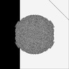 | |||||||||
|---|---|---|---|---|---|---|---|---|---|---|
| Title | Cryo-EM Structure of Human Enterovirus D68 USA/IL/14-18952 | |||||||||
 Map data Map data | Enterovirus D68 USA/IL/14-18952 apo | |||||||||
 Sample Sample |
| |||||||||
 Keywords Keywords | Virus / Enterovirus | |||||||||
| Function / homology |  Function and homology information Function and homology informationsymbiont-mediated suppression of cytoplasmic pattern recognition receptor signaling pathway / symbiont-mediated suppression of host cytoplasmic pattern recognition receptor signaling pathway via inhibition of MDA-5 activity / picornain 2A / symbiont-mediated suppression of host mRNA export from nucleus / symbiont genome entry into host cell via pore formation in plasma membrane / picornain 3C / protein sequestering activity / T=pseudo3 icosahedral viral capsid / host cell cytoplasmic vesicle membrane / ribonucleoside triphosphate phosphatase activity ...symbiont-mediated suppression of cytoplasmic pattern recognition receptor signaling pathway / symbiont-mediated suppression of host cytoplasmic pattern recognition receptor signaling pathway via inhibition of MDA-5 activity / picornain 2A / symbiont-mediated suppression of host mRNA export from nucleus / symbiont genome entry into host cell via pore formation in plasma membrane / picornain 3C / protein sequestering activity / T=pseudo3 icosahedral viral capsid / host cell cytoplasmic vesicle membrane / ribonucleoside triphosphate phosphatase activity / nucleoside-triphosphate phosphatase / channel activity / monoatomic ion transmembrane transport / symbiont-mediated suppression of host toll-like receptor signaling pathway / symbiont-mediated suppression of host NF-kappaB cascade / RNA helicase activity / symbiont-mediated suppression of host innate immune response / endocytosis involved in viral entry into host cell / symbiont-mediated activation of host autophagy / RNA-directed RNA polymerase / cysteine-type endopeptidase activity / viral RNA genome replication / RNA-directed RNA polymerase activity / virion attachment to host cell / host cell nucleus / DNA-templated transcription / structural molecule activity / proteolysis / RNA binding / zinc ion binding / ATP binding / cytoplasm Similarity search - Function | |||||||||
| Biological species |  enterovirus D68 enterovirus D68 | |||||||||
| Method | single particle reconstruction / cryo EM / Resolution: 2.0 Å | |||||||||
 Authors Authors | Xu L / Pintilie G / Varanese L / Carette JE / Chiu W | |||||||||
| Funding support |  United States, 1 items United States, 1 items
| |||||||||
 Citation Citation | Journal: Nature / Year: 2025 Title: MFSD6 is an entry receptor for enterovirus D68. Authors: Lauren Varanese / Lily Xu / Christine E Peters / Grigore Pintilie / David S Roberts / Suyash Raj / Mengying Liu / Yaw Shin Ooi / Jonathan Diep / Wenjie Qiao / Christopher M Richards / Jeremy ...Authors: Lauren Varanese / Lily Xu / Christine E Peters / Grigore Pintilie / David S Roberts / Suyash Raj / Mengying Liu / Yaw Shin Ooi / Jonathan Diep / Wenjie Qiao / Christopher M Richards / Jeremy Callaway / Carolyn R Bertozzi / Sabrina Jabs / Erik de Vries / Frank J M van Kuppeveld / Claude M Nagamine / Wah Chiu / Jan E Carette /    Abstract: With the near eradication of poliovirus due to global vaccination campaigns, attention has shifted to other enteroviruses that can cause polio-like paralysis syndrome (now termed acute flaccid ...With the near eradication of poliovirus due to global vaccination campaigns, attention has shifted to other enteroviruses that can cause polio-like paralysis syndrome (now termed acute flaccid myelitis). In particular, enterovirus D68 (EV-D68) is believed to be the main driver of epidemic outbreaks of acute flaccid myelitis in recent years, yet not much is known about EV-D68 host interactions. EV-D68 is a respiratory virus but, in rare cases, can spread to the central nervous system to cause severe neuropathogenesis. Here we use genome-scale CRISPR screens to identify the poorly characterized multipass membrane transporter MFSD6 as a host entry factor for EV-D68. Knockout of MFSD6 expression abrogated EV-D68 infection in cell lines and primary cells corresponding to respiratory and neural cells. MFSD6 localized to the plasma membrane and was required for viral entry into host cells. MFSD6 bound directly to EV-D68 particles through its extracellular, third loop (L3). We determined the cryo-electron microscopy structure of EV-D68 in a complex with MFSD6 L3, revealing the interaction interface. A decoy receptor, engineered by fusing MFSD6 L3 to Fc, blocked EV-D68 infection of human primary lung epithelial cells and provided near-complete protection in a lethal mouse model of EV-D68 infection. Collectively, our results reveal MFSD6 as an entry receptor for EV-D68, and support the targeting of MFSD6 as a potential mechanism to combat infections by this emerging pathogen with pandemic potential. | |||||||||
| History |
|
- Structure visualization
| Supplemental images |
|---|

- Downloads & links
-EMDB archive
| Map data | emd_48705.map.gz | 836.8 MB | EMDB map data format | |
|---|---|---|---|---|
| Header (meta data) | emd-48705-v30.xmlemd-48705.xml | 27.4 KB 27.4 KB | Display Display | EMDB header |
| FSC (resolution estimation) | emd_48705_fsc.xml | 23.6 KB | Display | FSC data file |
| Images |  emd_48705.png emd_48705.png | 258.5 KB | ||
| Filedesc metadata | emd-48705.cif.gz | 7.3 KB | ||
| Others | emd_48705_half_map_1.map.gzemd_48705_half_map_2.map.gz | 1.3 GB 1.3 GB | ||
| Archive directory |  http://ftp.pdbj.org/pub/emdb/structures/EMD-48705ftp://ftp.pdbj.org/pub/emdb/structures/EMD-48705 http://ftp.pdbj.org/pub/emdb/structures/EMD-48705ftp://ftp.pdbj.org/pub/emdb/structures/EMD-48705 | HTTPS FTP |
-Related structure data
| Related structure data | 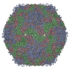 9mwzMC 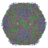 9mxcC C: citing same article ( M: atomic model generated by this map |
|---|---|
| Similar structure data |
-Links
| EMDB pages | EMDB (EBI/PDBe) / EMDataResource |
|---|---|
| Related items in Molecule of the Month |


-Map
| File | Download / File: emd_48705.map.gz / Format: CCP4 / Size: 1.4 GB / Type: IMAGE STORED AS FLOATING POINT NUMBER (4 BYTES) | ||||||||||||||||||||||||||||||||||||
|---|---|---|---|---|---|---|---|---|---|---|---|---|---|---|---|---|---|---|---|---|---|---|---|---|---|---|---|---|---|---|---|---|---|---|---|---|---|
| Annotation | Enterovirus D68 USA/IL/14-18952 apo | ||||||||||||||||||||||||||||||||||||
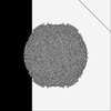
| Projections & slices | Image control
Images are generated by Spider. | ||||||||||||||||||||||||||||||||||||
| Voxel size | X=Y=Z: 0.743 Å | ||||||||||||||||||||||||||||||||||||
| Density |
| ||||||||||||||||||||||||||||||||||||
| Symmetry | Space group: 1 | ||||||||||||||||||||||||||||||||||||
| Details | EMDB XML:
|
 X (Sec.)
 Y (Row.)
 Z (Col.)


-Supplemental data
-Half map: Enterovirus D68 USA/IL/14-18952 apo half map A
| File | emd_48705_half_map_1.map | ||||||||||||
|---|---|---|---|---|---|---|---|---|---|---|---|---|---|
| Annotation | Enterovirus D68 USA/IL/14-18952 apo half map A | ||||||||||||
| Projections & Slices |
| ||||||||||||
| Density Histograms |


-Half map: Enterovirus D68 USA/IL/14-18952 apo half map B
| File | emd_48705_half_map_2.map | ||||||||||||
|---|---|---|---|---|---|---|---|---|---|---|---|---|---|
| Annotation | Enterovirus D68 USA/IL/14-18952 apo half map B | ||||||||||||
| Projections & Slices |
| ||||||||||||
| Density Histograms |


- Sample components
-Entire : enterovirus D68
| Entire | Name: enterovirus D68 |
|---|---|
| Components |
|
-Supramolecule #1: enterovirus D68
| Supramolecule | Name: enterovirus D68 / type: virus / ID: 1 / Parent: 0 / Macromolecule list: #1-#4 Details: Enterovirus D68 propagated and purified from infection of RD cells NCBI-ID: 42789 / Sci species name: enterovirus D68 / Sci species strain: 14-18952 / Virus type: VIRION / Virus isolate: STRAIN / Virus enveloped: No / Virus empty: No |
|---|---|
| Host (natural) | Organism:  Homo sapiens (human) Homo sapiens (human) |
-Macromolecule #1: viral protein 1
| Macromolecule | Name: viral protein 1 / type: protein_or_peptide / ID: 1 / Number of copies: 1 / Enantiomer: LEVO / EC number: picornain 2A |
|---|---|
| Source (natural) | Organism: enterovirus D68 / Strain: USA/IL/14-18952 |
| Molecular weight | Theoretical: 32.854242 KDa |
| Sequence | String: IESIIKTATD TVKSEINAEL GVVPSLNAVE TGATSNTEPE EAIQTRTVIN QHGVSETLVE NFLSRAALVS KRSFEYKDHT SSAAQTDKN FFKWTINTRS FVQLRRKLEL FTYLRFDAEI TILTTVAVNG SSNNTYVGLP DLTLQAMFVP TGALTPEKQD S FHWQSGSN ...String: IESIIKTATD TVKSEINAEL GVVPSLNAVE TGATSNTEPE EAIQTRTVIN QHGVSETLVE NFLSRAALVS KRSFEYKDHT SSAAQTDKN FFKWTINTRS FVQLRRKLEL FTYLRFDAEI TILTTVAVNG SSNNTYVGLP DLTLQAMFVP TGALTPEKQD S FHWQSGSN ASVFFKISDP PARMTIPFMC INSAYSVFYD GFAGFEKSGL YGINPADTIG NLCVRIVNEH QPVGFTVTVR VY MKPKHIK AWAPRPPRTL PYMSIANANY KGKGRAPNAL NAIIGNRDSV KTMPHNIVTT UniProtKB: Genome polyprotein |
-Macromolecule #2: viral protein 2
| Macromolecule | Name: viral protein 2 / type: protein_or_peptide / ID: 2 / Details: Residues 1-11 deleted / Number of copies: 1 / Enantiomer: LEVO / EC number: picornain 2A |
|---|---|
| Source (natural) | Organism: enterovirus D68 / Strain: USA/IL/14-18952 |
| Molecular weight | Theoretical: 26.527137 KDa |
| Sequence | String: RVLQLKLGNS AIVTQEAANY CCAYGEWPNY LPDHEAVAID KPTQPETATD RFYTLRSVKW EAGSTGWWWK LPDALNNIGM FGQNVQHHY LYRSGFLIHV QCNATKFHQG ALLVVAIPEH QRGAHNTNTS PGFDDIMKGE EGGTFNHPYV LDDGTSLACA T IFPHQWIN ...String: RVLQLKLGNS AIVTQEAANY CCAYGEWPNY LPDHEAVAID KPTQPETATD RFYTLRSVKW EAGSTGWWWK LPDALNNIGM FGQNVQHHY LYRSGFLIHV QCNATKFHQG ALLVVAIPEH QRGAHNTNTS PGFDDIMKGE EGGTFNHPYV LDDGTSLACA T IFPHQWIN LRTNNSATIV LPWMNAAPMD FPLRHNQWTL AIIPVVPLGT RTMSSMVPIT VSIAPMCCEF NGLRHAITQ UniProtKB: Genome polyprotein |
-Macromolecule #3: viral protein 3
| Macromolecule | Name: viral protein 3 / type: protein_or_peptide / ID: 3 / Number of copies: 1 / Enantiomer: LEVO / EC number: picornain 2A |
|---|---|
| Source (natural) | Organism: enterovirus D68 / Strain: USA/IL/14-18952 |
| Molecular weight | Theoretical: 27.156867 KDa |
| Sequence | String: GVPTYLLPGS GQFLTTDDHS SAPVLPCFNP TPEMHIPGQV RNMLEVVQVE SMMEINNTES AVGMERLKVD ISALTDVDQL LFNIPLDIQ LDGPLRNTLV GNISRYYTHW SGSLEMTFMF CGSFMATGKL ILCYTPPGGS CPTTRETAML GTHVVWDFGL Q SSVTLIIP ...String: GVPTYLLPGS GQFLTTDDHS SAPVLPCFNP TPEMHIPGQV RNMLEVVQVE SMMEINNTES AVGMERLKVD ISALTDVDQL LFNIPLDIQ LDGPLRNTLV GNISRYYTHW SGSLEMTFMF CGSFMATGKL ILCYTPPGGS CPTTRETAML GTHVVWDFGL Q SSVTLIIP WISGSHYRMF NNDAKSTNAN VGYVTCFMQT NLIVPSESSD TCSLIGFIAA KDDFSLRLMR DSPDIGQLDH LH AAEAAYQ UniProtKB: Genome polyprotein |
-Macromolecule #4: viral protein 4
| Macromolecule | Name: viral protein 4 / type: protein_or_peptide / ID: 4 / Details: Residues 1-27 and 58-68 deleted / Number of copies: 1 / Enantiomer: LEVO |
|---|---|
| Source (natural) | Organism: enterovirus D68 / Strain: USA/IL/14-18952 |
| Molecular weight | Theoretical: 3.401665 KDa |
| Sequence | String: QINFYKDSYA ASASKQDFSQ DPSKFTEPVV UniProtKB: Genome polyprotein |
-Macromolecule #5: water
| Macromolecule | Name: water / type: ligand / ID: 5 / Number of copies: 46 / Formula: HOH |
|---|---|
| Molecular weight | Theoretical: 18.015 Da |
| Chemical component information |  ChemComp-HOH: |
-Experimental details
-Structure determination
| Method | cryo EM |
|---|---|
 Processing Processing | single particle reconstruction |
| Aggregation state | particle |
-Sample preparation
| Concentration | 0.089 mg/mL | ||||||||||||
|---|---|---|---|---|---|---|---|---|---|---|---|---|---|
| Buffer | pH: 8 Component:
Details: 20 mM Tris-HCl (pH 8.0), 120 mM NaCl, and 1 mM EDTA (pH 8.0) | ||||||||||||
| Grid | Model: Quantifoil / Material: COPPER / Mesh: 400 / Support film - Material: CARBON / Support film - topology: LACEY / Support film - Film thickness: 3 / Pretreatment - Type: GLOW DISCHARGE / Pretreatment - Time: 30 sec. / Pretreatment - Atmosphere: AIR | ||||||||||||
| Vitrification | Cryogen name: ETHANE / Chamber humidity: 100 % / Chamber temperature: 277.15 K / Instrument: FEI VITROBOT MARK IV Details: Freezing carried out with 4 s blot time, 1 s wait time.. |
- Electron microscopy
| Microscope | TFS KRIOS |
|---|---|
| Image recording | Film or detector model: FEI FALCON IV (4k x 4k) / Number grids imaged: 1 / Number real images: 10400 / Average exposure time: 3.28 sec. / Average electron dose: 50.0 e/Å2 |
| Electron beam | Acceleration voltage: 300 kV / Electron source:  FIELD EMISSION GUN FIELD EMISSION GUN |
| Electron optics | Illumination mode: FLOOD BEAM / Imaging mode: DARK FIELD / Cs: 2.7 mm / Nominal defocus max: 2.0 µm / Nominal defocus min: 1.0 µm / Nominal magnification: 165000 |
| Sample stage | Specimen holder model: FEI TITAN KRIOS AUTOGRID HOLDER / Cooling holder cryogen: NITROGEN |
| Experimental equipment |  Model: Titan Krios / Image courtesy: FEI Company |
+Image processing

-Atomic model buiding 1
| Initial model | Chain - Source name: AlphaFold / Chain - Initial model type: in silico model |
|---|---|
| Details | Initial rigid fitting was done using Chimera and then ISOLDE was used for flexible fitting. |
| Refinement | Protocol: FLEXIBLE FIT |
| Output model | PDB-9mwz: |
