Movie
Movie Controller


+ Open data

- Basic information
| Entry |  | |||||||||||||||||||||||||||
|---|---|---|---|---|---|---|---|---|---|---|---|---|---|---|---|---|---|---|---|---|---|---|---|---|---|---|---|---|
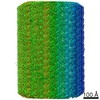
| Title | Doublet Microtubule from T. vaginalis - local refine 29 | |||||||||||||||||||||||||||
 Map data Map data | ||||||||||||||||||||||||||||
 Sample Sample |
| |||||||||||||||||||||||||||
 Keywords Keywords | microtubule / flagella / STRUCTURAL PROTEIN | |||||||||||||||||||||||||||
| Biological species |  Trichomonas vaginalis G3 (eukaryote) Trichomonas vaginalis G3 (eukaryote) | |||||||||||||||||||||||||||
| Method | single particle reconstruction / cryo EM / Resolution: 3.9 Å | |||||||||||||||||||||||||||
 Authors Authors | Stevens A / Zhou ZH | |||||||||||||||||||||||||||
| Funding support |  United States, 8 items United States, 8 items
| |||||||||||||||||||||||||||
 Citation Citation | Journal: To Be Published Title: Structures of Native Doublet Microtubules from Trichomonas vaginalis Reveal Parasite-Specific Proteins as Potential Drug Targets Authors: Stevens A / Saarang K / Crofut EC / Wang SE / Muratore KA / Johnson PJ / Zhou ZH | |||||||||||||||||||||||||||
| History |
|
- Structure visualization
| Supplemental images |
|---|

- Downloads & links
-EMDB archive
| Map data | emd_46027.map.gz | 943.1 MB |  EMDB map data format EMDB map data format | |
|---|---|---|---|---|
| Header (meta data) | emd-46027-v30.xmlemd-46027.xml | 20.6 KB 20.6 KB | Display Display | EMDB header |
| FSC (resolution estimation) | emd_46027_fsc.xml | 21.2 KB | Display | FSC data file |
| Images |  emd_46027.png emd_46027.png | 166.8 KB | ||
| Masks | emd_46027_msk_1.map | 1000 MB | Mask map | |
| Filedesc metadata | emd-46027.cif.gz | 4.9 KB | ||
| Others | emd_46027_half_map_1.map.gzemd_46027_half_map_2.map.gz | 929.1 MB 929.1 MB | ||
| Archive directory |  http://ftp.pdbj.org/pub/emdb/structures/EMD-46027ftp://ftp.pdbj.org/pub/emdb/structures/EMD-46027 http://ftp.pdbj.org/pub/emdb/structures/EMD-46027ftp://ftp.pdbj.org/pub/emdb/structures/EMD-46027 | HTTPS FTP |
-Validation report
| Summary document | emd_46027_validation.pdf.gz | 1.4 MB | Display | EMDB validaton report |
|---|---|---|---|---|
| Full document | emd_46027_full_validation.pdf.gz | 1.4 MB | Display | |
| Data in XML | emd_46027_validation.xml.gz | 30.6 KB | Display | |
| Data in CIF | emd_46027_validation.cif.gz | 40.7 KB | Display | |
| Arichive directory | https://ftp.pdbj.org/pub/emdb/validation_reports/EMD-46027ftp://ftp.pdbj.org/pub/emdb/validation_reports/EMD-46027 | HTTPS FTP |
-Related structure data
| Related structure data | C: citing same article ( |
|---|


-Links
| EMDB pages | EMDB (EBI/PDBe) / EMDataResource |
|---|
-Map
| File | Download / File: emd_46027.map.gz / Format: CCP4 / Size: 1000 MB / Type: IMAGE STORED AS FLOATING POINT NUMBER (4 BYTES) | ||||||||||||||||||||||||||||||||||||
|---|---|---|---|---|---|---|---|---|---|---|---|---|---|---|---|---|---|---|---|---|---|---|---|---|---|---|---|---|---|---|---|---|---|---|---|---|---|
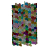
| Projections & slices | Image control
Images are generated by Spider. | ||||||||||||||||||||||||||||||||||||
| Voxel size | X=Y=Z: 1.1 Å | ||||||||||||||||||||||||||||||||||||
| Density |
| ||||||||||||||||||||||||||||||||||||
| Symmetry | Space group: 1 | ||||||||||||||||||||||||||||||||||||
| Details | EMDB XML:
|
 Z (Sec.)
 Y (Row.)
 X (Col.)


-Supplemental data
-Mask #1
| File | emd_46027_msk_1.map | ||||||||||||
|---|---|---|---|---|---|---|---|---|---|---|---|---|---|
| Projections & Slices |
| ||||||||||||
| Density Histograms |


-Half map: #1
| File | emd_46027_half_map_1.map | ||||||||||||
|---|---|---|---|---|---|---|---|---|---|---|---|---|---|
| Projections & Slices |
| ||||||||||||
| Density Histograms |


-Half map: #2
| File | emd_46027_half_map_2.map | ||||||||||||
|---|---|---|---|---|---|---|---|---|---|---|---|---|---|
| Projections & Slices |
| ||||||||||||
| Density Histograms |


- Sample components
-Entire : Flagellar doublet microtubules
| Entire | Name: Flagellar doublet microtubules |
|---|---|
| Components |
|
-Supramolecule #1: Flagellar doublet microtubules
| Supramolecule | Name: Flagellar doublet microtubules / type: complex / ID: 1 / Parent: 0 |
|---|---|
| Source (natural) | Organism: Trichomonas vaginalis G3 (eukaryote) / Location in cell: Flagella |
-Experimental details
-Structure determination
| Method | cryo EM |
|---|---|
 Processing Processing | single particle reconstruction |
| Aggregation state | particle |
-Sample preparation
| Buffer | pH: 7.4 Component:
Details: 150 mM NaCl, 50 mM 325 Tris, 2 mM MgCl2, 1 mM DTT, 5 mM ATP 2 x complete protease inhibitor, pH 7.4 | ||||||||||||||||||
|---|---|---|---|---|---|---|---|---|---|---|---|---|---|---|---|---|---|---|---|
| Grid | Model: Quantifoil R2/1 / Material: COPPER / Mesh: 200 / Support film - Material: CARBON / Support film - topology: HOLEY / Pretreatment - Type: GLOW DISCHARGE / Pretreatment - Time: 30 sec. | ||||||||||||||||||
| Vitrification | Cryogen name: ETHANE-PROPANE / Chamber humidity: 100 % / Chamber temperature: 281 K / Instrument: FEI VITROBOT MARK IV | ||||||||||||||||||
| Details | Doublet microtubules purified from T. vaginalis flagella |
- Electron microscopy
| Microscope | FEI TITAN |
|---|---|
| Image recording | Film or detector model: GATAN K3 (6k x 4k) / Number grids imaged: 1 / Average electron dose: 45.0 e/Å2 |
| Electron beam | Acceleration voltage: 300 kV / Electron source:  FIELD EMISSION GUN FIELD EMISSION GUN |
| Electron optics | Illumination mode: FLOOD BEAM / Imaging mode: BRIGHT FIELD / Cs: 2.7 mm / Nominal defocus max: 2.0 µm / Nominal defocus min: 1.2 µm / Nominal magnification: 81000 |
| Sample stage | Specimen holder model: FEI TITAN KRIOS AUTOGRID HOLDER / Cooling holder cryogen: NITROGEN |