 ムービー
ムービー コントローラー


+ データを開く

- 基本情報
基本情報
| 登録情報 | データベース: EMDB / ID: EMD-4602 | |||||||||
|---|---|---|---|---|---|---|---|---|---|---|
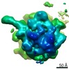
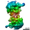
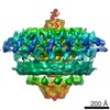
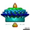
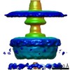
| タイトル | In situ Volta phase plate subtomogram average of phycobilisome array from Synechocystis | |||||||||
 マップデータ マップデータ | In situ bin4 Volta phase plate subtomogram average of phycobilisome array from Synechocystis sp. PCC 6803 | |||||||||
 試料 試料 |
| |||||||||
| 生物種 |  | |||||||||
| 手法 | サブトモグラム平均法 / クライオ電子顕微鏡法 / 解像度: 30.0 Å | |||||||||
 データ登録者 データ登録者 | Rast A / Wan W / Pfeffer S / Engel BD | |||||||||
| 資金援助 |  ドイツ, 1件 ドイツ, 1件
| |||||||||
 引用 引用 | ジャーナル: Nat Plants / 年: 2019 タイトル: Biogenic regions of cyanobacterial thylakoids form contact sites with the plasma membrane. 著者: Anna Rast / Miroslava Schaffer / Sahradha Albert / William Wan / Stefan Pfeffer / Florian Beck / Jürgen M Plitzko / Jörg Nickelsen / Benjamin D Engel / 要旨: Little is known about how the photosynthetic machinery is arranged in time and space during the biogenesis of thylakoid membranes. Using in situ cryo-electron tomography to image the three- ...Little is known about how the photosynthetic machinery is arranged in time and space during the biogenesis of thylakoid membranes. Using in situ cryo-electron tomography to image the three-dimensional architecture of the cyanobacterium Synechocystis, we observed that the tips of multiple thylakoids merge to form a substructure called the 'convergence membrane'. This high-curvature membrane comes into close contact with the plasma membrane at discrete sites. We generated subtomogram averages of 70S ribosomes and array-forming phycobilisomes, then mapped these structures onto the native membrane architecture as markers for protein synthesis and photosynthesis, respectively. This molecular localization identified two distinct biogenic regions in the thylakoid network: thylakoids facing the cytosolic interior of the cell that were associated with both marker complexes, and convergence membranes that were decorated by ribosomes but not phycobilisomes. We propose that the convergence membranes perform a specialized biogenic function, coupling the synthesis of thylakoid proteins with the integration of cofactors from the plasma membrane and the periplasmic space. | |||||||||
| 履歴 |
|
- 構造の表示
構造の表示
| ムービー |
 ムービービューア ムービービューア |
|---|---|
| 構造ビューア | EMマップ: SurfViewMolmilJmol/JSmol |
| 添付画像 |

- ダウンロードとリンク
-EMDBアーカイブ
| マップデータ | emd_4602.map.gz | 643.9 KB | EMDBマップデータ形式 | |
|---|---|---|---|---|
| ヘッダ (付随情報) | emd-4602-v30.xmlemd-4602.xml | 11.4 KB 11.4 KB | 表示 表示 | EMDBヘッダ |
| 画像 |  emd_4602.png emd_4602.png | 232.2 KB | ||
| アーカイブディレクトリ |  http://ftp.pdbj.org/pub/emdb/structures/EMD-4602ftp://ftp.pdbj.org/pub/emdb/structures/EMD-4602 http://ftp.pdbj.org/pub/emdb/structures/EMD-4602ftp://ftp.pdbj.org/pub/emdb/structures/EMD-4602 | HTTPS FTP |
-検証レポート
| 文書・要旨 | emd_4602_validation.pdf.gz | 247.9 KB | 表示 | EMDB検証レポート |
|---|---|---|---|---|
| 文書・詳細版 | emd_4602_full_validation.pdf.gz | 247 KB | 表示 | |
| XML形式データ | emd_4602_validation.xml.gz | 5.1 KB | 表示 | |
| アーカイブディレクトリ | https://ftp.pdbj.org/pub/emdb/validation_reports/EMD-4602ftp://ftp.pdbj.org/pub/emdb/validation_reports/EMD-4602 | HTTPS FTP |
-関連構造データ


-リンク
| EMDBのページ | EMDB (EBI/PDBe) / EMDataResource |
|---|
-マップ
| ファイル | ダウンロード / ファイル: emd_4602.map.gz / 形式: CCP4 / 大きさ: 686.5 KB / タイプ: IMAGE STORED AS FLOATING POINT NUMBER (4 BYTES) | ||||||||||||||||||||||||||||||||||||||||||||||||||||||||||||||||||||
|---|---|---|---|---|---|---|---|---|---|---|---|---|---|---|---|---|---|---|---|---|---|---|---|---|---|---|---|---|---|---|---|---|---|---|---|---|---|---|---|---|---|---|---|---|---|---|---|---|---|---|---|---|---|---|---|---|---|---|---|---|---|---|---|---|---|---|---|---|---|
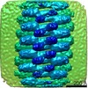
| 注釈 | In situ bin4 Volta phase plate subtomogram average of phycobilisome array from Synechocystis sp. PCC 6803 | ||||||||||||||||||||||||||||||||||||||||||||||||||||||||||||||||||||
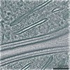
| 投影像・断面図 | 画像のコントロール
画像は Spider により作成 | ||||||||||||||||||||||||||||||||||||||||||||||||||||||||||||||||||||
| ボクセルのサイズ | X=Y=Z: 13.68 Å | ||||||||||||||||||||||||||||||||||||||||||||||||||||||||||||||||||||
| 密度 |
| ||||||||||||||||||||||||||||||||||||||||||||||||||||||||||||||||||||
| 対称性 | 空間群: 1 | ||||||||||||||||||||||||||||||||||||||||||||||||||||||||||||||||||||
| 詳細 | EMDB XML:
CCP4マップ ヘッダ情報:
| ||||||||||||||||||||||||||||||||||||||||||||||||||||||||||||||||||||
 Z (Sec.)
 Y (Row.)
 X (Col.)


-添付データ
- 試料の構成要素
-全体 : Phycobilisome within the native cellular environment of Synechocystis
| 全体 | 名称: Phycobilisome within the native cellular environment of Synechocystis |
|---|---|
| 要素 |
|
-超分子 #1: Phycobilisome within the native cellular environment of Synechocystis
| 超分子 | 名称: Phycobilisome within the native cellular environment of Synechocystis タイプ: complex / ID: 1 / 親要素: 0 詳細: The molecular weight of a single unit within the array is ~6 MDa. |
|---|---|
| 由来(天然) | 生物種: |
| 分子量 | 理論値: 30 MDa |
-実験情報
-構造解析
| 手法 | クライオ電子顕微鏡法 |
|---|---|
 解析 解析 | サブトモグラム平均法 |
| 試料の集合状態 | cell |
-試料調製
| 緩衝液 | pH: 7 |
|---|---|
| グリッド | モデル: Quantifoil R1.2/1.3 / 材質: COPPER / メッシュ: 200 / 前処理 - タイプ: GLOW DISCHARGE / 前処理 - 雰囲気: AIR |
| 凍結 | 凍結剤: ETHANE-PROPANE / チャンバー内湿度: 90 % / チャンバー内温度: 297 K / 装置: FEI VITROBOT MARK IV / 詳細: Blotting time: 10 sec Blot force: 10. |
| 詳細 | Vitrious Synechocystis cell milled with a Ga2+ focused ion beam. |
- 電子顕微鏡法
| 顕微鏡 | FEI TITAN KRIOS |
|---|---|
| 特殊光学系 | 位相板: VOLTA PHASE PLATE / エネルギーフィルター - 名称: GIF Quantum LS / エネルギーフィルター - スリット幅: 20 eV |
| 撮影 | フィルム・検出器のモデル: GATAN K2 SUMMIT (4k x 4k) 検出モード: COUNTING / デジタル化 - サイズ - 横: 3838 pixel / デジタル化 - サイズ - 縦: 3710 pixel / 平均露光時間: 1.5 sec. / 平均電子線量: 1.5 e/Å2 詳細: Images were collected in movie-mode at 12 frames per second. Higher tilts had longer exposures. |
| 電子線 | 加速電圧: 300 kV / 電子線源:  FIELD EMISSION GUN FIELD EMISSION GUN |
| 電子光学系 | C2レンズ絞り径: 70.0 µm / 照射モード: FLOOD BEAM / 撮影モード: BRIGHT FIELD / Cs: 2.7 mm / 最大 デフォーカス(公称値): 0.0005 µm / 最小 デフォーカス(公称値): 0.0001 µm / 倍率(公称値): 42000 |
| 試料ステージ | 試料ホルダーモデル: FEI TITAN KRIOS AUTOGRID HOLDER ホルダー冷却材: NITROGEN |
| 実験機器 |  モデル: Titan Krios / 画像提供: FEI Company |
-画像解析
| 最終 再構成 | 想定した対称性 - 点群: C1 (非対称) / 解像度のタイプ: BY AUTHOR / 解像度: 30.0 Å / 解像度の算出法: FSC 0.5 CUT-OFF / ソフトウェア - 名称: PyTom (ver. 0.97) / 使用したサブトモグラム数: 600 |
|---|---|
| 抽出 | トモグラム数: 5 / 使用した粒子像数: 1000 / ソフトウェア - 名称: PyTom (ver. 0.97) 詳細: Template matching in PyTom with a de novo reference. |
| 最終 角度割当 | タイプ: OTHER / ソフトウェア - 名称: PyTom (ver. 0.97) / 詳細: template matching |
