 Movie
Movie Controller

[English] 日本語
 Yorodumi
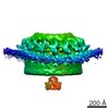
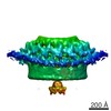
- EMDB-4601: In situ defocus subtomogram average of phycobilisome array from S... -
+ Open data

- Basic information
| Entry | Database: EMDB / ID: EMD-4601 | |||||||||
|---|---|---|---|---|---|---|---|---|---|---|
| Title | In situ defocus subtomogram average of phycobilisome array from Synechocystis sp. PCC 6803 | |||||||||
 Map data Map data | In situ defocus subtomogram average of phycobilisome array from Synechocystis sp. PCC 6803 | |||||||||
 Sample Sample |
| |||||||||
| Biological species |  | |||||||||
| Method | subtomogram averaging / cryo EM / Resolution: 23.6 Å | |||||||||
 Authors Authors | Rast A / Wan W / Pfeffer S / Engel BD | |||||||||
 Citation Citation | Journal: Nat Plants / Year: 2019 Title: Biogenic regions of cyanobacterial thylakoids form contact sites with the plasma membrane. Authors: Anna Rast / Miroslava Schaffer / Sahradha Albert / William Wan / Stefan Pfeffer / Florian Beck / Jürgen M Plitzko / Jörg Nickelsen / Benjamin D Engel /  Abstract: Little is known about how the photosynthetic machinery is arranged in time and space during the biogenesis of thylakoid membranes. Using in situ cryo-electron tomography to image the three- ...Little is known about how the photosynthetic machinery is arranged in time and space during the biogenesis of thylakoid membranes. Using in situ cryo-electron tomography to image the three-dimensional architecture of the cyanobacterium Synechocystis, we observed that the tips of multiple thylakoids merge to form a substructure called the 'convergence membrane'. This high-curvature membrane comes into close contact with the plasma membrane at discrete sites. We generated subtomogram averages of 70S ribosomes and array-forming phycobilisomes, then mapped these structures onto the native membrane architecture as markers for protein synthesis and photosynthesis, respectively. This molecular localization identified two distinct biogenic regions in the thylakoid network: thylakoids facing the cytosolic interior of the cell that were associated with both marker complexes, and convergence membranes that were decorated by ribosomes but not phycobilisomes. We propose that the convergence membranes perform a specialized biogenic function, coupling the synthesis of thylakoid proteins with the integration of cofactors from the plasma membrane and the periplasmic space. | |||||||||
| History |
|
- Structure visualization
| Movie |
 Movie viewer Movie viewer |
|---|---|
| Structure viewer | EM map: SurfViewMolmilJmol/JSmol |
| Supplemental images |

- Downloads & links
-EMDB archive
| Map data | emd_4601.map.gz | 58 MB | EMDB map data format | |
|---|---|---|---|---|
| Header (meta data) | emd-4601-v30.xmlemd-4601.xml | 11 KB 11 KB | Display Display | EMDB header |
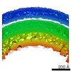
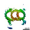
| Images | 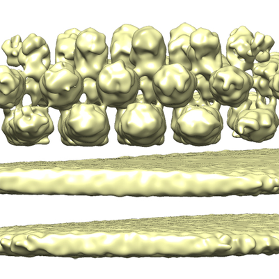 emd_4601.png emd_4601.png | 256.8 KB | ||
| Archive directory |  http://ftp.pdbj.org/pub/emdb/structures/EMD-4601ftp://ftp.pdbj.org/pub/emdb/structures/EMD-4601 http://ftp.pdbj.org/pub/emdb/structures/EMD-4601ftp://ftp.pdbj.org/pub/emdb/structures/EMD-4601 | HTTPS FTP |
-Related structure data

-Links
| EMDB pages | EMDB (EBI/PDBe) / EMDataResource |
|---|
-Map
| File | Download / File: emd_4601.map.gz / Format: CCP4 / Size: 64 MB / Type: IMAGE STORED AS FLOATING POINT NUMBER (4 BYTES) | ||||||||||||||||||||||||||||||||||||||||||||||||||||||||||||
|---|---|---|---|---|---|---|---|---|---|---|---|---|---|---|---|---|---|---|---|---|---|---|---|---|---|---|---|---|---|---|---|---|---|---|---|---|---|---|---|---|---|---|---|---|---|---|---|---|---|---|---|---|---|---|---|---|---|---|---|---|---|
| Annotation | In situ defocus subtomogram average of phycobilisome array from Synechocystis sp. PCC 6803 | ||||||||||||||||||||||||||||||||||||||||||||||||||||||||||||
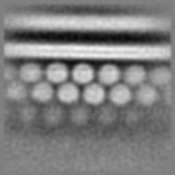
| Projections & slices | Image control
Images are generated by Spider. | ||||||||||||||||||||||||||||||||||||||||||||||||||||||||||||
| Voxel size | X=Y=Z: 3.42 Å | ||||||||||||||||||||||||||||||||||||||||||||||||||||||||||||
| Density |
| ||||||||||||||||||||||||||||||||||||||||||||||||||||||||||||
| Symmetry | Space group: 1 | ||||||||||||||||||||||||||||||||||||||||||||||||||||||||||||
| Details | EMDB XML:
CCP4 map header:
| ||||||||||||||||||||||||||||||||||||||||||||||||||||||||||||
 Z (Sec.)
 Y (Row.)
 X (Col.)


-Supplemental data
- Sample components
-Entire : In situ defocus subtomogram average of phycobilisome array from S...
| Entire | Name: In situ defocus subtomogram average of phycobilisome array from Synechocystis |
|---|---|
| Components |
|
-Supramolecule #1: In situ defocus subtomogram average of phycobilisome array from S...
| Supramolecule | Name: In situ defocus subtomogram average of phycobilisome array from Synechocystis type: complex / ID: 1 / Parent: 0 Details: Each unit of the array has a molecular weight of ~6 MDa. |
|---|---|
| Source (natural) | Organism: |
| Molecular weight | Theoretical: 30 MDa |
-Experimental details
-Structure determination
| Method | cryo EM |
|---|---|
 Processing Processing | subtomogram averaging |
| Aggregation state | cell |
-Sample preparation
| Buffer | pH: 7 |
|---|---|
| Grid | Model: Quantifoil R1.2/1.3 / Material: COPPER / Mesh: 200 / Pretreatment - Type: GLOW DISCHARGE / Pretreatment - Atmosphere: AIR |
| Vitrification | Cryogen name: ETHANE-PROPANE / Chamber humidity: 90 % / Chamber temperature: 297 K / Instrument: FEI VITROBOT MARK IV / Details: Blotting time: 10 sec Blot force: 10. |
| Details | Vitrious Synechocystis cell milled with a Ga2+ focused ion beam. |
- Electron microscopy
| Microscope | FEI TITAN KRIOS |
|---|---|
| Specialist optics | Energy filter - Name: GIF Quantum LS / Energy filter - Slit width: 20 eV |
| Image recording | Film or detector model: GATAN K2 SUMMIT (4k x 4k) / Detector mode: COUNTING / Digitization - Dimensions - Width: 3838 pixel / Digitization - Dimensions - Height: 3710 pixel / Average exposure time: 1.5 sec. / Average electron dose: 1.5 e/Å2 Details: Images were collected in movie-mode at 12 frames per second. Higher tilts had longer exposures. |
| Electron beam | Acceleration voltage: 300 kV / Electron source:  FIELD EMISSION GUN FIELD EMISSION GUN |
| Electron optics | C2 aperture diameter: 70.0 µm / Illumination mode: FLOOD BEAM / Imaging mode: BRIGHT FIELD / Cs: 2.7 mm / Nominal defocus max: 0.006 µm / Nominal defocus min: 0.004 µm / Nominal magnification: 42000 |
| Sample stage | Specimen holder model: FEI TITAN KRIOS AUTOGRID HOLDER / Cooling holder cryogen: NITROGEN |
| Experimental equipment |  Model: Titan Krios / Image courtesy: FEI Company |
-Image processing
| Final reconstruction | Applied symmetry - Point group: C2 (2 fold cyclic) / Resolution.type: BY AUTHOR / Resolution: 23.6 Å / Resolution method: FSC 0.143 CUT-OFF / Details: Averaging was performed with STOPGAP 0.3.1 / Number subtomograms used: 1710 |
|---|---|
| Extraction | Number tomograms: 20 / Number images used: 3850 / Software - Name: PyTom (ver. 0.97) Details: Template matching followed by oversampling of splines along arrays. Duplicate points were removed following the alignment process. |
| CTF correction | Software - Name: IMOD (ver. 4.9) |
| Final angle assignment | Type: OTHER / Details: Template matching with PyTom |
