 Movie
Movie Controller


+ Open data

- Basic information
| Entry |  | |||||||||
|---|---|---|---|---|---|---|---|---|---|---|
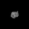
| Title | Open state SARS-COV2 spike protein from EMPIAR-10453 | |||||||||
 Map data Map data | ||||||||||
 Sample Sample |
| |||||||||
 Keywords Keywords | ribosome / tomography / 70S | |||||||||
| Biological species |   Severe acute respiratory syndrome coronavirus 2 Severe acute respiratory syndrome coronavirus 2 | |||||||||
| Method | subtomogram averaging / cryo EM / Resolution: 5.6 Å | |||||||||
 Authors Authors | Zhou Y / Huang QW / Bartesaghi A | |||||||||
| Funding support |  United States, 1 items United States, 1 items
| |||||||||
 Citation Citation | Journal: Nat Methods / Year: 2024 Title: MiLoPYP: self-supervised molecular pattern mining and particle localization in situ. Authors: Qinwen Huang / Ye Zhou / Alberto Bartesaghi / Abstract: Cryo-electron tomography allows the routine visualization of cellular landscapes in three dimensions at nanometer-range resolutions. When combined with single-particle tomography, it is possible to ...Cryo-electron tomography allows the routine visualization of cellular landscapes in three dimensions at nanometer-range resolutions. When combined with single-particle tomography, it is possible to obtain near-atomic resolution structures of frequently occurring macromolecules within their native environment. Two outstanding challenges associated with cryo-electron tomography/single-particle tomography are the automatic identification and localization of proteins, tasks that are hindered by the molecular crowding inside cells, imaging distortions characteristic of cryo-electron tomography tomograms and the sheer size of tomographic datasets. Current methods suffer from low accuracy, demand extensive and time-consuming manual labeling or are limited to the detection of specific types of proteins. Here, we present MiLoPYP, a two-step dataset-specific contrastive learning-based framework that enables fast molecular pattern mining followed by accurate protein localization. MiLoPYP's ability to effectively detect and localize a wide range of targets including globular and tubular complexes as well as large membrane proteins, will contribute to streamline and broaden the applicability of high-resolution workflows for in situ structure determination. | |||||||||
| History |
|
- Structure visualization
| Supplemental images |
|---|

- Downloads & links
-EMDB archive
| Map data | emd_45267.map.gz | 810.1 KB |  EMDB map data format EMDB map data format | |
|---|---|---|---|---|
| Header (meta data) | emd-45267-v30.xmlemd-45267.xml | 11.5 KB 11.5 KB | Display Display | EMDB header |
| Images |  emd_45267.png emd_45267.png | 77.9 KB | ||
| Masks | emd_45267_msk_1.map | 10.5 MB | Mask map | |
| Filedesc metadata | emd-45267.cif.gz | 3.8 KB | ||
| Others | emd_45267_half_map_1.map.gzemd_45267_half_map_2.map.gz | 5.6 MB 5.6 MB | ||
| Archive directory |  http://ftp.pdbj.org/pub/emdb/structures/EMD-45267ftp://ftp.pdbj.org/pub/emdb/structures/EMD-45267 http://ftp.pdbj.org/pub/emdb/structures/EMD-45267ftp://ftp.pdbj.org/pub/emdb/structures/EMD-45267 | HTTPS FTP |
-Related structure data


-Links
| EMDB pages | EMDB (EBI/PDBe) / EMDataResource |
|---|
-Map
| File | Download / File: emd_45267.map.gz / Format: CCP4 / Size: 10.5 MB / Type: IMAGE STORED AS FLOATING POINT NUMBER (4 BYTES) | ||||||||||||||||||||||||||||||||||||
|---|---|---|---|---|---|---|---|---|---|---|---|---|---|---|---|---|---|---|---|---|---|---|---|---|---|---|---|---|---|---|---|---|---|---|---|---|---|
| Projections & slices | Image control
Images are generated by Spider. | ||||||||||||||||||||||||||||||||||||
| Voxel size | X=Y=Z: 2.658 Å | ||||||||||||||||||||||||||||||||||||
| Density |
| ||||||||||||||||||||||||||||||||||||
| Symmetry | Space group: 1 | ||||||||||||||||||||||||||||||||||||
| Details | EMDB XML:
|
 Z (Sec.)
 Y (Row.)
 X (Col.)


-Supplemental data
-Mask #1
| File | emd_45267_msk_1.map | ||||||||||||
|---|---|---|---|---|---|---|---|---|---|---|---|---|---|
| Projections & Slices |
| ||||||||||||
| Density Histograms |


-Half map: #1
| File | emd_45267_half_map_1.map | ||||||||||||
|---|---|---|---|---|---|---|---|---|---|---|---|---|---|
| Projections & Slices |
| ||||||||||||
| Density Histograms |


-Half map: #2
| File | emd_45267_half_map_2.map | ||||||||||||
|---|---|---|---|---|---|---|---|---|---|---|---|---|---|
| Projections & Slices |
| ||||||||||||
| Density Histograms |


- Sample components
-Entire : Severe acute respiratory syndrome coronavirus 2
| Entire | Name: Severe acute respiratory syndrome coronavirus 2 |
|---|---|
| Components |
|
-Supramolecule #1: Severe acute respiratory syndrome coronavirus 2
| Supramolecule | Name: Severe acute respiratory syndrome coronavirus 2 / type: virus / ID: 1 / Parent: 0 / NCBI-ID: 2697049 Sci species name: Severe acute respiratory syndrome coronavirus 2 Virus type: VIRION / Virus isolate: STRAIN / Virus enveloped: No / Virus empty: No |
|---|
-Experimental details
-Structure determination
| Method | cryo EM |
|---|---|
 Processing Processing | subtomogram averaging |
| Aggregation state | cell |
-Sample preparation
| Buffer | pH: 7.4 |
|---|---|
| Vitrification | Cryogen name: ETHANE |
- Electron microscopy
| Microscope | FEI TITAN KRIOS |
|---|---|
| Image recording | Film or detector model: GATAN K3 (6k x 4k) / Average electron dose: 3.5 e/Å2 |
| Electron beam | Acceleration voltage: 300 kV / Electron source:  FIELD EMISSION GUN FIELD EMISSION GUN |
| Electron optics | Illumination mode: FLOOD BEAM / Imaging mode: BRIGHT FIELD / Nominal defocus max: 3.5 µm / Nominal defocus min: 1.5 µm |
| Experimental equipment |  Model: Titan Krios / Image courtesy: FEI Company |
-Image processing
| Final reconstruction | Resolution.type: BY AUTHOR / Resolution: 5.6 Å / Resolution method: FSC 0.143 CUT-OFF / Number subtomograms used: 9194 |
|---|---|
| Extraction | Number tomograms: 266 / Number images used: 23388 |
| Final angle assignment | Type: MAXIMUM LIKELIHOOD |
