Journal: Nat Methods / Year: 2024 Title: MiLoPYP: self-supervised molecular pattern mining and particle localization in situ. Authors: Qinwen Huang / Ye Zhou / Alberto Bartesaghi / Abstract: Cryo-electron tomography allows the routine visualization of cellular landscapes in three dimensions at nanometer-range resolutions. When combined with single-particle tomography, it is possible to ...Cryo-electron tomography allows the routine visualization of cellular landscapes in three dimensions at nanometer-range resolutions. When combined with single-particle tomography, it is possible to obtain near-atomic resolution structures of frequently occurring macromolecules within their native environment. Two outstanding challenges associated with cryo-electron tomography/single-particle tomography are the automatic identification and localization of proteins, tasks that are hindered by the molecular crowding inside cells, imaging distortions characteristic of cryo-electron tomography tomograms and the sheer size of tomographic datasets. Current methods suffer from low accuracy, demand extensive and time-consuming manual labeling or are limited to the detection of specific types of proteins. Here, we present MiLoPYP, a two-step dataset-specific contrastive learning-based framework that enables fast molecular pattern mining followed by accurate protein localization. MiLoPYP's ability to effectively detect and localize a wide range of targets including globular and tubular complexes as well as large membrane proteins, will contribute to streamline and broaden the applicability of high-resolution workflows for in situ structure determination.
In the structure databanks used in Yorodumi, some data are registered as the other names, "COVID-19 virus" and "2019-nCoV". Here are the details of the virus and the list of structure data.
Jan 31, 2019. EMDB accession codes are about to change! (news from PDBe EMDB page)
EMDB accession codes are about to change! (news from PDBe EMDB page)
The allocation of 4 digits for EMDB accession codes will soon come to an end. Whilst these codes will remain in use, new EMDB accession codes will include an additional digit and will expand incrementally as the available range of codes is exhausted. The current 4-digit format prefixed with “EMD-” (i.e. EMD-XXXX) will advance to a 5-digit format (i.e. EMD-XXXXX), and so on. It is currently estimated that the 4-digit codes will be depleted around Spring 2019, at which point the 5-digit format will come into force.
The EM Navigator/Yorodumi systems omit the EMD- prefix.
Related info.:Q: What is EMD? / ID/Accession-code notation in Yorodumi/EM Navigator
Yorodumi is a browser for structure data from EMDB, PDB, SASBDB, etc.
This page is also the successor to EM Navigator detail page, and also detail information page/front-end page for Omokage search.
The word "yorodu" (or yorozu) is an old Japanese word meaning "ten thousand". "mi" (miru) is to see.
Related info.:EMDB / PDB / SASBDB / Comparison of 3 databanks / Yorodumi Search / Aug 31, 2016. New EM Navigator & Yorodumi / Yorodumi Papers / Jmol/JSmol / Function and homology information / Changes in new EM Navigator and Yorodumi
 Movie
Movie Controller


 Open data
 Basic information
 Map data
 Sample
 Keywords
 Authors
 United States, 1 items
  Citation
 Structure visualization
 Downloads & links
 EMDB map data format
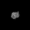
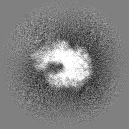
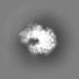
 emd_45261.png
 http://ftp.pdbj.org/pub/emdb/structures/EMD-45261


 Z (Sec.)
 Y (Row.)
 X (Col.)


 Sample components
 Processing
 Electron microscopy
 FIELD EMISSION GUN
