 Movie
Movie Controller
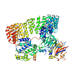


+ Open data

- Basic information
| Entry |  | |||||||||
|---|---|---|---|---|---|---|---|---|---|---|
| Title | PP2A:B55-p107 substrate complex | |||||||||
 Map data Map data | Primary Map NU-Refinment, Cryosparc, phenix | |||||||||
 Sample Sample |
| |||||||||
 Keywords Keywords | PP2A:B55 / p107 / substrate complex / HYDROLASE / HYDROLASE-SUBSTRATE complex | |||||||||
| Function / homology |  Function and homology information Function and homology informationregulation of lipid kinase activity / regulation of chromosome segregation / meiotic spindle elongation / Integration of energy metabolism / PP2A-mediated dephosphorylation of key metabolic factors / RNA polymerase II CTD heptapeptide repeat S2 phosphatase activity / RNA polymerase II CTD heptapeptide repeat S7 phosphatase activity / mitotic sister chromatid separation / MASTL Facilitates Mitotic Progression / protein phosphatase type 2A complex ...regulation of lipid kinase activity / regulation of chromosome segregation / meiotic spindle elongation / Integration of energy metabolism / PP2A-mediated dephosphorylation of key metabolic factors / RNA polymerase II CTD heptapeptide repeat S2 phosphatase activity / RNA polymerase II CTD heptapeptide repeat S7 phosphatase activity / mitotic sister chromatid separation / MASTL Facilitates Mitotic Progression / protein phosphatase type 2A complex / protein serine/threonine phosphatase complex / regulation of meiotic cell cycle process involved in oocyte maturation / peptidyl-threonine dephosphorylation / meiotic sister chromatid cohesion, centromeric / INTAC complex / RNA polymerase II CTD heptapeptide repeat S5 phosphatase activity / FAR/SIN/STRIPAK complex / Regulation of glycolysis by fructose 2,6-bisphosphate metabolism / Inhibition of replication initiation of damaged DNA by RB1/E2F1 / female meiotic nuclear division / protein phosphatase regulator activity / GABA receptor binding / APC truncation mutants have impaired AXIN binding / AXIN missense mutants destabilize the destruction complex / Truncations of AMER1 destabilize the destruction complex / Transcription of E2F targets under negative control by p107 (RBL1) and p130 (RBL2) in complex with HDAC1 / protein antigen binding / ERKs are inactivated / Initiation of Nuclear Envelope (NE) Reformation / positive regulation of extrinsic apoptotic signaling pathway in absence of ligand / Beta-catenin phosphorylation cascade / Signaling by GSK3beta mutants / CTNNB1 S33 mutants aren't phosphorylated / CTNNB1 S37 mutants aren't phosphorylated / CTNNB1 S45 mutants aren't phosphorylated / CTNNB1 T41 mutants aren't phosphorylated / RNA polymerase II transcription initiation surveillance / Transcription of E2F targets under negative control by DREAM complex / Co-stimulation by CD28 / regulation of growth / Disassembly of the destruction complex and recruitment of AXIN to the membrane / negative regulation of epithelial to mesenchymal transition / response to morphine / Co-inhibition by CTLA4 / Platelet sensitization by LDL / negative regulation of G1/S transition of mitotic cell cycle / protein-serine/threonine phosphatase / negative regulation of glycolytic process through fructose-6-phosphate / positive regulation of NLRP3 inflammasome complex assembly / ERK/MAPK targets / G1/S-Specific Transcription / mesoderm development / protein serine/threonine phosphatase activity / vascular endothelial cell response to oscillatory fluid shear stress / T cell homeostasis / regulation of cell differentiation / regulation of G1/S transition of mitotic cell cycle / regulation of microtubule polymerization / phosphoprotein phosphatase activity / negative regulation of cellular senescence / G0 and Early G1 / lateral plasma membrane / chromosome, centromeric region / DARPP-32 events / enzyme-substrate adaptor activity / negative regulation of hippo signaling / Nonsense Mediated Decay (NMD) enhanced by the Exon Junction Complex (EJC) / Cyclin A/B1/B2 associated events during G2/M transition / protein dephosphorylation / spindle assembly / Amplification of signal from unattached kinetochores via a MAD2 inhibitory signal / Mitotic Prometaphase / EML4 and NUDC in mitotic spindle formation / Loss of Nlp from mitotic centrosomes / Loss of proteins required for interphase microtubule organization from the centrosome / Recruitment of mitotic centrosome proteins and complexes / negative regulation of phosphatidylinositol 3-kinase/protein kinase B signal transduction / Recruitment of NuMA to mitotic centrosomes / Anchoring of the basal body to the plasma membrane / protein tyrosine phosphatase activity / Resolution of Sister Chromatid Cohesion / TP53 Regulates Transcription of Genes Involved in G2 Cell Cycle Arrest / protein phosphatase 2A binding / AURKA Activation by TPX2 / Turbulent (oscillatory, disturbed) flow shear stress activates signaling by PIEZO1 and integrins in endothelial cells / SMAD2/SMAD3:SMAD4 heterotrimer regulates transcription / Spry regulation of FGF signaling / meiotic cell cycle / chromosome segregation / RNA polymerase II transcription regulatory region sequence-specific DNA binding / Degradation of beta-catenin by the destruction complex / promoter-specific chromatin binding / RHO GTPases Activate Formins / RAF activation / negative regulation of canonical Wnt signaling pathway / PKR-mediated signaling / Negative regulation of MAPK pathway / response to lead ion / tau protein binding / Cyclin D associated events in G1 Similarity search - Function | |||||||||
| Biological species |  Homo sapiens (human) Homo sapiens (human) | |||||||||
| Method | single particle reconstruction / cryo EM / Resolution: 2.6 Å | |||||||||
 Authors Authors | Page R / Peti W / Padi S / Godek RJ | |||||||||
| Funding support |  United States, 2 items United States, 2 items
| |||||||||
 Citation Citation | Journal: Nat.Struct.Mol.Biol. / Year: 2025 Title: Cryo-EM structures of PP2A:B55 with p107 and Eya3 define substrate recruitment. Authors: Padi SKR / Godek RJ / Peti W / Page R | |||||||||
| History |
|
- Structure visualization
- Downloads & links
-EMDB archive
| Map data | emd_45243.map.gz | 72.7 MB | EMDB map data format | |
|---|---|---|---|---|
| Header (meta data) | emd-45243-v30.xmlemd-45243.xml | 23.8 KB 23.8 KB | Display Display | EMDB header |
| FSC (resolution estimation) | emd_45243_fsc.xml | 12.4 KB | Display | FSC data file |
| Images |  emd_45243.png emd_45243.png | 86.1 KB | ||
| Masks | emd_45243_msk_1.map | 144.7 MB | Mask map | |
| Filedesc metadata | emd-45243.cif.gz | 7.3 KB | ||
| Others | emd_45243_half_map_1.map.gzemd_45243_half_map_2.map.gz | 134.2 MB 134.2 MB | ||
| Archive directory |  http://ftp.pdbj.org/pub/emdb/structures/EMD-45243ftp://ftp.pdbj.org/pub/emdb/structures/EMD-45243 http://ftp.pdbj.org/pub/emdb/structures/EMD-45243ftp://ftp.pdbj.org/pub/emdb/structures/EMD-45243 | HTTPS FTP |
-Validation report
| Summary document | emd_45243_validation.pdf.gz | 893.1 KB | Display | EMDB validaton report |
|---|---|---|---|---|
| Full document | emd_45243_full_validation.pdf.gz | 892.7 KB | Display | |
| Data in XML | emd_45243_validation.xml.gz | 19.8 KB | Display | |
| Data in CIF | emd_45243_validation.cif.gz | 25.3 KB | Display | |
| Arichive directory | https://ftp.pdbj.org/pub/emdb/validation_reports/EMD-45243ftp://ftp.pdbj.org/pub/emdb/validation_reports/EMD-45243 | HTTPS FTP |
-Related structure data
| Related structure data |  9c6bMC  9c7tC M: atomic model generated by this map C: citing same article ( |
|---|---|
| Similar structure data |

-Links
| EMDB pages | EMDB (EBI/PDBe) / EMDataResource |
|---|---|
| Related items in Molecule of the Month |


-Map
| File | Download / File: emd_45243.map.gz / Format: CCP4 / Size: 144.7 MB / Type: IMAGE STORED AS FLOATING POINT NUMBER (4 BYTES) | ||||||||||||||||||||
|---|---|---|---|---|---|---|---|---|---|---|---|---|---|---|---|---|---|---|---|---|---|
| Annotation | Primary Map NU-Refinment, Cryosparc, phenix | ||||||||||||||||||||
| Voxel size | X=Y=Z: 0.8266 Å | ||||||||||||||||||||
| Density |
| ||||||||||||||||||||
| Symmetry | Space group: 1 | ||||||||||||||||||||
| Details | EMDB XML:
|
-Supplemental data
- Sample components
-Entire : PP2A:B55-p107
| Entire | Name: PP2A:B55-p107 |
|---|---|
| Components |
|
-Supramolecule #1: PP2A:B55-p107
| Supramolecule | Name: PP2A:B55-p107 / type: complex / ID: 1 / Parent: 0 / Macromolecule list: #1-#4 Details: The PP2A:B55 complex (PP2Aa:B55:PP2Ac) bound to p107 |
|---|---|
| Molecular weight | Theoretical: 161.4 KDa |
-Macromolecule #1: Serine/threonine-protein phosphatase 2A 65 kDa regulatory subunit...
| Macromolecule | Name: Serine/threonine-protein phosphatase 2A 65 kDa regulatory subunit A alpha isoform type: protein_or_peptide / ID: 1 / Number of copies: 1 / Enantiomer: LEVO |
|---|---|
| Source (natural) | Organism: Homo sapiens (human) |
| Molecular weight | Theoretical: 64.95798 KDa |
| Recombinant expression | Organism:  |
| Sequence | String: GHMSLYPIAV LIDELRNEDV QLRLNSIKKL STIALALGVE RTRSELLPFL TDTIYDEDEV LLALAEQLGT FTTLVGGPEY VHCLLPPLE SLATVEETVV RDKAVESLRA ISHEHSPSDL EAHFVPLVKR LAGGDWFTSR TSACGLFSVC YPRVSSAVKA E LRQYFRNL ...String: GHMSLYPIAV LIDELRNEDV QLRLNSIKKL STIALALGVE RTRSELLPFL TDTIYDEDEV LLALAEQLGT FTTLVGGPEY VHCLLPPLE SLATVEETVV RDKAVESLRA ISHEHSPSDL EAHFVPLVKR LAGGDWFTSR TSACGLFSVC YPRVSSAVKA E LRQYFRNL CSDDTPMVRR AAASKLGEFA KVLELDNVKS EIIPMFSNLA SDEQDSVRLL AVEACVNIAQ LLPQEDLEAL VM PTLRQAA EDKSWRVRYM VADKFTELQK AVGPEITKTD LVPAFQNLMK DCEAEVRAAA SHKVKEFCEN LSADCRENVI MSQ ILPCIK ELVSDANQHV KSALASVIMG LSPILGKDNT IEHLLPLFLA QLKDECPEVR LNIISNLDCV NEVIGIRQLS QSLL PAIVE LAEDAKWRVR LAIIEYMPLL AGQLGVEFFD EKLNSLCMAW LVDHVYAIRE AATSNLKKLV EKFGKEWAHA TIIPK VLAM SGDPNYLHRM TTLFCINVLS EVCGQDITTK HMLPTVLRMA GDPVANVRFN VAKSLQKIGP ILDNSTLQSE VKPILE KLT QDQDVDVKYF AQEALTVLSL A UniProtKB: Serine/threonine-protein phosphatase 2A 65 kDa regulatory subunit A alpha isoform |
-Macromolecule #2: Serine/threonine-protein phosphatase 2A 55 kDa regulatory subunit...
| Macromolecule | Name: Serine/threonine-protein phosphatase 2A 55 kDa regulatory subunit B alpha isoform type: protein_or_peptide / ID: 2 / Number of copies: 1 / Enantiomer: LEVO |
|---|---|
| Source (natural) | Organism: Homo sapiens (human) |
| Molecular weight | Theoretical: 52.10134 KDa |
| Recombinant expression | Organism: Homo sapiens (human) |
| Sequence | String: GHMGSAGAGG GNDIQWCFSQ VKGAVDDDVA EADIISTVEF NHSGELLATG DKGGRVVIFQ QEQENKIQSH SRGEYNVYST FQSHEPEFD YLKSLEIEEK INKIRWLPQK NAAQFLLSTN DKTIKLWKIS ERDKRPEGYN LKEEDGRYRD PTTVTTLRVP V FRPMDLMV ...String: GHMGSAGAGG GNDIQWCFSQ VKGAVDDDVA EADIISTVEF NHSGELLATG DKGGRVVIFQ QEQENKIQSH SRGEYNVYST FQSHEPEFD YLKSLEIEEK INKIRWLPQK NAAQFLLSTN DKTIKLWKIS ERDKRPEGYN LKEEDGRYRD PTTVTTLRVP V FRPMDLMV EASPRRIFAN AHTYHINSIS INSDYETYLS ADDLRINLWH LEITDRSFNI VDIKPANMEE LTEVITAAEF HP NSCNTFV YSSSKGTIRL CDMRASALCD RHSKLFEEPE DPSNRSFFSE IISSISDVKF SHSGRYMMTR DYLSVKIWDL NME NRPVET YQVHEYLRSK LCSLYENDCI FDKFECCWNG SDSVVMTGSY NNFFRMFDRN TKRDITLEAS RENNKPRTVL KPRK VCASG KRKKDEISVD SLDFNKKILH TAWHPKENII AVATTNNLYI FQDKVN UniProtKB: Serine/threonine-protein phosphatase 2A 55 kDa regulatory subunit B alpha isoform |
-Macromolecule #3: Serine/threonine-protein phosphatase 2A catalytic subunit alpha i...
| Macromolecule | Name: Serine/threonine-protein phosphatase 2A catalytic subunit alpha isoform type: protein_or_peptide / ID: 3 / Number of copies: 1 / Enantiomer: LEVO / EC number: protein-serine/threonine phosphatase |
|---|---|
| Source (natural) | Organism: Homo sapiens (human) |
| Molecular weight | Theoretical: 35.845375 KDa |
| Recombinant expression | Organism: Homo sapiens (human) |
| Sequence | String: GHMDEKVFTK ELDQWIEQLN ECKQLSESQV KSLCEKAKEI LTKESNVQEV RCPVTVCGDV HGQFHDLMEL FRIGGKSPDT NYLFMGDYV DRGYYSVETV TLLVALKVRY RERITILRGN HESRQITQVY GFYDECLRKY GNANVWKYFT DLFDYLPLTA L VDGQIFCL ...String: GHMDEKVFTK ELDQWIEQLN ECKQLSESQV KSLCEKAKEI LTKESNVQEV RCPVTVCGDV HGQFHDLMEL FRIGGKSPDT NYLFMGDYV DRGYYSVETV TLLVALKVRY RERITILRGN HESRQITQVY GFYDECLRKY GNANVWKYFT DLFDYLPLTA L VDGQIFCL HGGLSPSIDT LDHIRALDRL QEVPHEGPMC DLLWSDPDDR GGWGISPRGA GYTFGQDISE TFNHANGLTL VS RAHQLVM EGYNWCHDRN VVTIFSAPNY CYRCGNQAAI MELDDTLKYS FLQFDPAPRR GEPHVTRRTP DYF(MLL) UniProtKB: Serine/threonine-protein phosphatase 2A catalytic subunit alpha isoform |
-Macromolecule #4: Retinoblastoma-like protein 1
| Macromolecule | Name: Retinoblastoma-like protein 1 / type: protein_or_peptide / ID: 4 / Number of copies: 1 / Enantiomer: LEVO |
|---|---|
| Source (natural) | Organism: Homo sapiens (human) |
| Molecular weight | Theoretical: 8.716149 KDa |
| Recombinant expression | Organism: |
| Sequence | String: GHMPMSPLMH PRVKEVRTDS GSLRRDMQPL SPISVHERYS SPTAGSAKRR LFGEDPPKEM LMDKIITEGT KLKIAPSS UniProtKB: Retinoblastoma-like protein 1 |
-Macromolecule #5: FE (II) ION
| Macromolecule | Name: FE (II) ION / type: ligand / ID: 5 / Number of copies: 1 / Formula: FE2 |
|---|---|
| Molecular weight | Theoretical: 55.845 Da |
-Macromolecule #6: ZINC ION
| Macromolecule | Name: ZINC ION / type: ligand / ID: 6 / Number of copies: 1 / Formula: ZN |
|---|---|
| Molecular weight | Theoretical: 65.409 Da |
-Experimental details
-Structure determination
| Method | cryo EM |
|---|---|
 Processing Processing | single particle reconstruction |
| Aggregation state | particle |
-Sample preparation
| Buffer | pH: 8 |
|---|---|
| Vitrification | Cryogen name: ETHANE |
- Electron microscopy
| Microscope | FEI TITAN KRIOS |
|---|---|
| Image recording | Film or detector model: GATAN K3 BIOCONTINUUM (6k x 4k) / Number grids imaged: 1 / Number real images: 5007 / Average electron dose: 39.6 e/Å2 |
| Electron beam | Acceleration voltage: 300 kV / Electron source:  FIELD EMISSION GUN FIELD EMISSION GUN |
| Electron optics | Illumination mode: FLOOD BEAM / Imaging mode: BRIGHT FIELD / Nominal defocus max: 2.6 µm / Nominal defocus min: 0.55 µm |
| Experimental equipment |  Model: Titan Krios / Image courtesy: FEI Company |
+Image processing
-Atomic model buiding 1
| Refinement | Space: REAL / Protocol: FLEXIBLE FIT / Overall B value: 60 / Target criteria: Cross-correlation coefficient |
|---|---|
| Output model | PDB-9c6b: |
