 Movie
Movie Controller


+ Open data

- Basic information
| Entry | Database: PDB / ID: 9c6b | |||||||||
|---|---|---|---|---|---|---|---|---|---|---|
| Title | PP2A:B55-p107 substrate complex | |||||||||
 Components Components |
| |||||||||
 Keywords Keywords | HYDROLASE/SUBSTRATE / PP2A:B55 / p107 / substrate complex / HYDROLASE / HYDROLASE-SUBSTRATE complex | |||||||||
| Function / homology |  Function and homology information Function and homology informationregulation of lipid kinase activity / regulation of chromosome segregation / meiotic spindle elongation / PP2A-mediated dephosphorylation of key metabolic factors / RNA polymerase II CTD heptapeptide repeat S2 phosphatase activity / RNA polymerase II CTD heptapeptide repeat S7 phosphatase activity / peptidyl-threonine dephosphorylation / regulation of meiotic cell cycle process involved in oocyte maturation / mitotic sister chromatid separation / MASTL Facilitates Mitotic Progression ...regulation of lipid kinase activity / regulation of chromosome segregation / meiotic spindle elongation / PP2A-mediated dephosphorylation of key metabolic factors / RNA polymerase II CTD heptapeptide repeat S2 phosphatase activity / RNA polymerase II CTD heptapeptide repeat S7 phosphatase activity / peptidyl-threonine dephosphorylation / regulation of meiotic cell cycle process involved in oocyte maturation / mitotic sister chromatid separation / MASTL Facilitates Mitotic Progression / protein phosphatase type 2A complex / meiotic sister chromatid cohesion, centromeric / INTAC complex / RNA polymerase II CTD heptapeptide repeat S5 phosphatase activity / FAR/SIN/STRIPAK complex / female meiotic nuclear division / Regulation of glycolysis by fructose 2,6-bisphosphate metabolism / Inhibition of replication initiation of damaged DNA by RB1/E2F1 / protein phosphatase regulator activity / protein antigen binding / Transcription of E2F targets under negative control by p107 (RBL1) and p130 (RBL2) in complex with HDAC1 / GABA receptor binding / APC truncation mutants have impaired AXIN binding / AXIN missense mutants destabilize the destruction complex / Truncations of AMER1 destabilize the destruction complex / positive regulation of extrinsic apoptotic signaling pathway in absence of ligand / ERKs are inactivated / Initiation of Nuclear Envelope (NE) Reformation / Beta-catenin phosphorylation cascade / Signaling by GSK3beta mutants / CTNNB1 S33 mutants aren't phosphorylated / CTNNB1 S37 mutants aren't phosphorylated / CTNNB1 S45 mutants aren't phosphorylated / CTNNB1 T41 mutants aren't phosphorylated / Transcription of E2F targets under negative control by DREAM complex / Co-stimulation by CD28 / regulation of growth / RNA polymerase II transcription initiation surveillance / Disassembly of the destruction complex and recruitment of AXIN to the membrane / protein dephosphorylation / negative regulation of epithelial to mesenchymal transition / Co-inhibition by CTLA4 / Platelet sensitization by LDL / negative regulation of G1/S transition of mitotic cell cycle / protein-serine/threonine phosphatase / G1/S-Specific Transcription / response to morphine / ERK/MAPK targets / negative regulation of glycolytic process through fructose-6-phosphate / vascular endothelial cell response to oscillatory fluid shear stress / protein serine/threonine phosphatase activity / mesoderm development / T cell homeostasis / positive regulation of NLRP3 inflammasome complex assembly / negative regulation of cellular senescence / regulation of cell differentiation / regulation of microtubule polymerization / G0 and Early G1 / regulation of G1/S transition of mitotic cell cycle / lateral plasma membrane / chromosome, centromeric region / DARPP-32 events / enzyme-substrate adaptor activity / negative regulation of hippo signaling / Cyclin A/B1/B2 associated events during G2/M transition / Nonsense Mediated Decay (NMD) enhanced by the Exon Junction Complex (EJC) / spindle assembly / phosphoprotein phosphatase activity / Amplification of signal from unattached kinetochores via a MAD2 inhibitory signal / Loss of Nlp from mitotic centrosomes / Loss of proteins required for interphase microtubule organization from the centrosome / Recruitment of mitotic centrosome proteins and complexes / protein tyrosine phosphatase activity / Recruitment of NuMA to mitotic centrosomes / Anchoring of the basal body to the plasma membrane / Mitotic Prometaphase / TP53 Regulates Transcription of Genes Involved in G2 Cell Cycle Arrest / EML4 and NUDC in mitotic spindle formation / protein phosphatase 2A binding / Turbulent (oscillatory, disturbed) flow shear stress activates signaling by PIEZO1 and integrins in endothelial cells / AURKA Activation by TPX2 / Resolution of Sister Chromatid Cohesion / negative regulation of phosphatidylinositol 3-kinase/protein kinase B signal transduction / meiotic cell cycle / RNA polymerase II transcription regulatory region sequence-specific DNA binding / promoter-specific chromatin binding / chromosome segregation / SMAD2/SMAD3:SMAD4 heterotrimer regulates transcription / negative regulation of canonical Wnt signaling pathway / RAF activation / RHO GTPases Activate Formins / Spry regulation of FGF signaling / PKR-mediated signaling / response to lead ion / Degradation of beta-catenin by the destruction complex / tau protein binding / spindle pole / Cyclin D associated events in G1 / Negative regulation of MAPK pathway / Separation of Sister Chromatids Similarity search - Function | |||||||||
| Biological species |  Homo sapiens (human) Homo sapiens (human) | |||||||||
| Method | ELECTRON MICROSCOPY / single particle reconstruction / cryo EM / Resolution: 2.6 Å | |||||||||
 Authors Authors | Page, R. / Peti, W. / Padi, S. / Godek, R.J. | |||||||||
| Funding support |  United States, 2items United States, 2items
| |||||||||
 Citation Citation | Journal: Nat Struct Mol Biol / Year: 2025 Title: Cryo-EM structures of PP2A:B55 with p107 and Eya3 define substrate recruitment. Authors: Sathish K R Padi / Rachel J Godek / Wolfgang Peti / Rebecca Page / Abstract: Phosphoprotein phosphatases (PPPs) achieve specificity by binding substrates and regulators using PPP-specific short motifs. Protein phosphatase 2A (PP2A) is a highly conserved phosphatase that ...Phosphoprotein phosphatases (PPPs) achieve specificity by binding substrates and regulators using PPP-specific short motifs. Protein phosphatase 2A (PP2A) is a highly conserved phosphatase that regulates cell signaling and is a tumor suppressor. Here, we use cryo-electron microscopy and nuclear magnetic resonance (NMR) spectroscopy to investigate the mechanisms of human p107 substrate and Eya3 regulator recruitment to the PP2A:B55 holoenzyme. We show that, while they associate with B55 using a common set of interaction pockets, the mechanism of substrate and regulator binding differs and is distinct from that observed for PP2A:B56 and other PPPs. We also identify the core B55 recruitment motif in Eya3 proteins, a sequence conserved amongst the Eya family. Lastly, using NMR-based dephosphorylation assays, we demonstrate how B55 recruitment directs PP2A:B55 fidelity through the selective dephosphorylation of specific phosphosites. As PP2A:B55 orchestrates mitosis and DNA damage repair, these data provide a roadmap for pursuing new avenues to therapeutically target this complex by individually blocking a subset of regulators that use different B55 interaction sites. #1: Journal: Nature / Year: 2024Title: Cryo-EM structures of PP2A:B55-FAM122A and PP2A:B55-ARPP19. Authors: Sathish K R Padi / Margaret R Vos / Rachel J Godek / James R Fuller / Thomas Kruse / Jamin B Hein / Jakob Nilsson / Matthew S Kelker / Rebecca Page / Wolfgang Peti /  Abstract: Progression through the cell cycle is controlled by regulated and abrupt changes in phosphorylation. Mitotic entry is initiated by increased phosphorylation of mitotic proteins, a process driven by ...Progression through the cell cycle is controlled by regulated and abrupt changes in phosphorylation. Mitotic entry is initiated by increased phosphorylation of mitotic proteins, a process driven by kinases, whereas mitotic exit is achieved by counteracting dephosphorylation, a process driven by phosphatases, especially PP2A:B55. Although the role of kinases in mitotic entry is well established, recent data have shown that mitosis is only successfully initiated when the counterbalancing phosphatases are also inhibited. Inhibition of PP2A:B55 is achieved by the intrinsically disordered proteins ARPP19 and FAM122A. Despite their critical roles in mitosis, the mechanisms by which they achieve PP2A:B55 inhibition is unknown. Here, we report the single-particle cryo-electron microscopy structures of PP2A:B55 bound to phosphorylated ARPP19 and FAM122A. Consistent with our complementary NMR spectroscopy studies, both intrinsically disordered proteins bind PP2A:B55, but do so in highly distinct manners, leveraging multiple distinct binding sites on B55. Our extensive structural, biophysical and biochemical data explain how substrates and inhibitors are recruited to PP2A:B55 and provide a molecular roadmap for the development of therapeutic interventions for PP2A:B55-related diseases. | |||||||||
| History |
|
- Structure visualization
| Structure viewer | Molecule: MolmilJmol/JSmol |
|---|
- Downloads & links
-Download
| PDBx/mmCIF format | 9c6b.cif.gz | 252.1 KB | Display | PDBx/mmCIF format |
|---|---|---|---|---|
| PDB format | pdb9c6b.ent.gz | 195.6 KB | Display | PDB format |
| PDBx/mmJSON format | 9c6b.json.gz | Tree view | PDBx/mmJSON format | |
| Others |  Other downloads Other downloads |
-Validation report
| Arichive directory | https://data.pdbj.org/pub/pdb/validation_reports/c6/9c6bftp://data.pdbj.org/pub/pdb/validation_reports/c6/9c6b | HTTPS FTP |
|---|
-Related structure data
| Related structure data |  45243MC 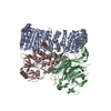 9c7tC M: map data used to model this data C: citing same article ( |
|---|---|
| Similar structure data |

-Links
 PDBj
PDBj


- Assembly
| Deposited unit | 
|
|---|---|
| 1 |
|
-Components
-Serine/threonine-protein phosphatase 2A ... , 3 types, 3 molecules ABC
| #1: Protein | Mass: 64957.980 Da / Num. of mol.: 1 / Fragment: residues 9-589 Source method: isolated from a genetically manipulated source Source: (gene. exp.) Homo sapiens (human) / Gene: PPP2R1A / Production host:  |
|---|---|
| #2: Protein | Mass: 52101.340 Da / Num. of mol.: 1 Source method: isolated from a genetically manipulated source Source: (gene. exp.) Homo sapiens (human) / Gene: PPP2R2A / Production host: Homo sapiens (human) / References: UniProt: P63151 |
| #3: Protein | Mass: 35845.375 Da / Num. of mol.: 1 Source method: isolated from a genetically manipulated source Source: (gene. exp.) Homo sapiens (human) / Gene: PPP2CA / Production host: Homo sapiens (human)References: UniProt: P67775, protein-serine/threonine phosphatase |
-Protein , 1 types, 1 molecules D
| #4: Protein | Mass: 8716.149 Da / Num. of mol.: 1 / Fragment: residues 612-687 Source method: isolated from a genetically manipulated source Source: (gene. exp.) Homo sapiens (human) / Gene: RBL1 / Production host: |
|---|
-Non-polymers , 2 types, 2 molecules 

| #5: Chemical | ChemComp-FE2 /  Mass: 55.845 Da / Num. of mol.: 1 / Source method: obtained synthetically / Formula: Fe Mass: 55.845 Da / Num. of mol.: 1 / Source method: obtained synthetically / Formula: Fe |
|---|---|
| #6: Chemical | ChemComp-ZN /  Mass: 65.409 Da / Num. of mol.: 1 / Source method: obtained synthetically / Formula: Zn Mass: 65.409 Da / Num. of mol.: 1 / Source method: obtained synthetically / Formula: Zn |
-Details
| Has ligand of interest | N |
|---|---|
| Has protein modification | Y |
-Experimental details
-Experiment
| Experiment | Method: ELECTRON MICROSCOPY |
|---|---|
| EM experiment | Aggregation state: PARTICLE / 3D reconstruction method: single particle reconstruction |
- Sample preparation
| Component | Name: PP2A:B55-p107 / Type: COMPLEX Details: The PP2A:B55 complex (PP2Aa:B55:PP2Ac) bound to p107 Entity ID: #1-#4 / Source: MULTIPLE SOURCES |
|---|---|
| Molecular weight | Value: 0.1614 MDa / Experimental value: NO |
| Buffer solution | pH: 8 |
| Specimen | Embedding applied: NO / Shadowing applied: NO / Staining applied: NO / Vitrification applied: YES |
| Vitrification | Cryogen name: ETHANE |
- Electron microscopy imaging
| Experimental equipment |  Model: Titan Krios / Image courtesy: FEI Company |
|---|---|
| Microscopy | Model: FEI TITAN KRIOS |
| Electron gun | Electron source:  FIELD EMISSION GUN / Accelerating voltage: 300 kV / Illumination mode: FLOOD BEAM FIELD EMISSION GUN / Accelerating voltage: 300 kV / Illumination mode: FLOOD BEAM |
| Electron lens | Mode: BRIGHT FIELD / Nominal defocus max: 2600 nm / Nominal defocus min: 550 nm |
| Image recording | Electron dose: 39.6 e/Å2 / Film or detector model: GATAN K3 BIOCONTINUUM (6k x 4k) / Num. of grids imaged: 1 / Num. of real images: 5007 |
- Processing
| EM software |
| ||||||||||||||||||||||||||||||||||||||||||||||||||||||||||||||||||||||||||||||||
|---|---|---|---|---|---|---|---|---|---|---|---|---|---|---|---|---|---|---|---|---|---|---|---|---|---|---|---|---|---|---|---|---|---|---|---|---|---|---|---|---|---|---|---|---|---|---|---|---|---|---|---|---|---|---|---|---|---|---|---|---|---|---|---|---|---|---|---|---|---|---|---|---|---|---|---|---|---|---|---|---|---|
| CTF correction | Type: PHASE FLIPPING AND AMPLITUDE CORRECTION | ||||||||||||||||||||||||||||||||||||||||||||||||||||||||||||||||||||||||||||||||
| Particle selection | Num. of particles selected: 2800783 | ||||||||||||||||||||||||||||||||||||||||||||||||||||||||||||||||||||||||||||||||
| 3D reconstruction | Resolution: 2.6 Å / Resolution method: FSC 0.143 CUT-OFF / Num. of particles: 195950 / Symmetry type: POINT | ||||||||||||||||||||||||||||||||||||||||||||||||||||||||||||||||||||||||||||||||
| Atomic model building | B value: 60 / Protocol: FLEXIBLE FIT / Space: REAL / Target criteria: Cross-correlation coefficient | ||||||||||||||||||||||||||||||||||||||||||||||||||||||||||||||||||||||||||||||||
| Refine LS restraints |
|
