National Institutes of Health/National Institute of General Medical Sciences (NIH/NIGMS)
R35 GM150960
米国
引用
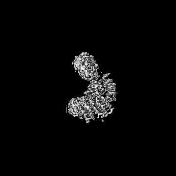
ジャーナル: Proc Natl Acad Sci U S A / 年: 2024 タイトル: A structure-based mechanism for initiation of AP-3 coated vesicle formation. 著者: Matthew Begley / Mahira Aragon / Richard W Baker / 要旨: Adaptor protein complex-3 (AP-3) mediates cargo sorting from endosomes to lysosomes and lysosome-related organelles. Recently, it was shown that AP-3 adopts a constitutively open conformation ...Adaptor protein complex-3 (AP-3) mediates cargo sorting from endosomes to lysosomes and lysosome-related organelles. Recently, it was shown that AP-3 adopts a constitutively open conformation compared to the related AP-1 and AP-2 coat complexes, which are inactive until undergoing large conformational changes upon membrane recruitment. How AP-3 is regulated is therefore an open question. To understand the mechanism of AP-3 membrane recruitment and activation, we reconstituted human AP-3 and determined multiple structures in the soluble and membrane-bound states using electron cryo-microscopy. Similar to yeast AP-3, human AP-3 is in a constitutively open conformation. To reconstitute AP-3 activation by adenosine di-phosphate (ADP)-ribosylation factor 1 (Arf1), a small guanosine tri-phosphate (GTP)ase, we used lipid nanodiscs to build Arf1-AP-3 complexes on membranes and determined three structures showing the stepwise conformational changes required for formation of AP-3 coated vesicles. First, membrane recruitment is driven by one of two predicted Arf1 binding sites, which flexibly tethers AP-3 to the membrane. Second, cargo binding causes AP-3 to adopt a fixed position and rigidifies the complex, which stabilizes binding for a second Arf1 molecule. Finally, binding of the second Arf1 molecule provides the template for AP-3 dimerization, providing a glimpse into the first step of coat polymerization. We propose coat polymerization only occurs after cargo engagement, thereby linking cargo sorting with assembly of higher-order coat structures. Additionally, we provide evidence for two amphipathic helices in AP-3, suggesting that AP-3 contributes to membrane deformation during coat assembly. In total, these data provide evidence for the first stages of AP-3-mediated vesicle coat assembly.
 ムービー
ムービー コントローラー


 データを開く
 基本情報
基本情報
 マップデータ
 試料
試料 キーワード
キーワード 機能・相同性情報
 Homo sapiens (ヒト)
 データ登録者
 米国, 1件
  引用
引用 構造の表示
構造の表示
 ダウンロードとリンク
 emd_45214.png
 http://ftp.pdbj.org/pub/emdb/structures/EMD-45214


 Z (Sec.)
 Y (Row.)
 X (Col.)


 試料の構成要素
 解析
解析 電子顕微鏡法
 FIELD EMISSION GUN


