 ムービー
ムービー コントローラー


+ データを開く

- 基本情報
基本情報
| 登録情報 |  | |||||||||
|---|---|---|---|---|---|---|---|---|---|---|
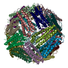
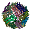
| タイトル | Human light chain ferritin reacted with iron (3 Fe2+ to ferritin monomer ratio). Reconstruction of particles with one nanoparticle. | |||||||||
 マップデータ マップデータ | Human light chain ferritin reacted with Ferrous salt(3 Fe2 per ferritin subunit) .Map based on 2D classes which show one iron nanoparticle per ferritin cage. | |||||||||
 試料 試料 |
| |||||||||
 キーワード キーワード | Iron oxide binding protein / METAL BINDING PROTEIN | |||||||||
| 機能・相同性 |  機能・相同性情報 機能・相同性情報ferritin complex / Scavenging by Class A Receptors / Golgi Associated Vesicle Biogenesis / autolysosome / ferric iron binding / autophagosome / Iron uptake and transport / iron ion transport / ferrous iron binding / azurophil granule lumen ...ferritin complex / Scavenging by Class A Receptors / Golgi Associated Vesicle Biogenesis / autolysosome / ferric iron binding / autophagosome / Iron uptake and transport / iron ion transport / ferrous iron binding / azurophil granule lumen / intracellular iron ion homeostasis / iron ion binding / Neutrophil degranulation / extracellular exosome / extracellular region / identical protein binding / membrane / cytosol / cytoplasm 類似検索 - 分子機能 | |||||||||
| 生物種 |  Homo sapiens (ヒト) Homo sapiens (ヒト) | |||||||||
| 手法 | 単粒子再構成法 / クライオ電子顕微鏡法 / 解像度: 2.85 Å | |||||||||
 データ登録者 データ登録者 | Sen S / Nannenga BL / Williams D | |||||||||
| 資金援助 |  米国, 1件 米国, 1件
| |||||||||
 引用 引用 | ジャーナル: J Am Chem Soc / 年: 2025 タイトル: Observation of the Protein-Inorganic Interface of Ferritin by Cryo-Electron Microscopy. 著者: Sagnik Sen / Amar Thaker / Alison Haymaker / Dewight Williams / Po-Lin Chiu / Brent L Nannenga / 要旨: Visualizing the structure of the protein-inorganic interface is critically important for a more complete understanding of biomineralization. Unfortunately, there are limited approaches for the direct ...Visualizing the structure of the protein-inorganic interface is critically important for a more complete understanding of biomineralization. Unfortunately, there are limited approaches for the direct and detailed study of biomolecules that interact with inorganic materials. Here, we use single-particle cryo-electron microscopy (cryo-EM) to study the protein-nanoparticle (NP) interactions of human light chain ferritin and visualize the high-resolution details of the protein-inorganic interface. In this work, we determined the 2.85 Å structure of human light chain ferritin bound to its native iron oxide NP substrate. The resulting cryo-EM maps confirmed and enhanced previously proposed interactions of the protein with the material along the B-helix and revealed new interaction at the C-terminus of light chain ferritin. This work sheds new light on the mechanisms of ferritin biomineralization and further demonstrates the application of cryo-EM for the study of protein-inorganic systems. | |||||||||
| 履歴 |
|
- 構造の表示
構造の表示
| 添付画像 |
|---|

- ダウンロードとリンク
-EMDBアーカイブ
| マップデータ | emd_44779.map.gz | 32.4 MB | EMDBマップデータ形式 | |
|---|---|---|---|---|
| ヘッダ (付随情報) | emd-44779-v30.xmlemd-44779.xml | 13.8 KB 13.8 KB | 表示 表示 | EMDBヘッダ |
| FSC (解像度算出) | emd_44779_fsc.xml | 8.4 KB | 表示 | FSCデータファイル |
| 画像 |  emd_44779.png emd_44779.png | 58 KB | ||
| Filedesc metadata | emd-44779.cif.gz | 5.3 KB | ||
| その他 | emd_44779_half_map_1.map.gzemd_44779_half_map_2.map.gz | 59.4 MB 59.4 MB | ||
| アーカイブディレクトリ |  http://ftp.pdbj.org/pub/emdb/structures/EMD-44779ftp://ftp.pdbj.org/pub/emdb/structures/EMD-44779 http://ftp.pdbj.org/pub/emdb/structures/EMD-44779ftp://ftp.pdbj.org/pub/emdb/structures/EMD-44779 | HTTPS FTP |
-検証レポート
| 文書・要旨 | emd_44779_validation.pdf.gz | 1.1 MB | 表示 | EMDB検証レポート |
|---|---|---|---|---|
| 文書・詳細版 | emd_44779_full_validation.pdf.gz | 1.1 MB | 表示 | |
| XML形式データ | emd_44779_validation.xml.gz | 16.5 KB | 表示 | |
| CIF形式データ | emd_44779_validation.cif.gz | 21.3 KB | 表示 | |
| アーカイブディレクトリ | https://ftp.pdbj.org/pub/emdb/validation_reports/EMD-44779ftp://ftp.pdbj.org/pub/emdb/validation_reports/EMD-44779 | HTTPS FTP |
-関連構造データ


-リンク
| EMDBのページ | EMDB (EBI/PDBe) / EMDataResource |
|---|---|
| 「今月の分子」の関連する項目 |


-マップ
| ファイル | ダウンロード / ファイル: emd_44779.map.gz / 形式: CCP4 / 大きさ: 64 MB / タイプ: IMAGE STORED AS FLOATING POINT NUMBER (4 BYTES) | ||||||||||||||||||||||||||||||||||||
|---|---|---|---|---|---|---|---|---|---|---|---|---|---|---|---|---|---|---|---|---|---|---|---|---|---|---|---|---|---|---|---|---|---|---|---|---|---|
| 注釈 | Human light chain ferritin reacted with Ferrous salt(3 Fe2 per ferritin subunit) .Map based on 2D classes which show one iron nanoparticle per ferritin cage. | ||||||||||||||||||||||||||||||||||||
| 投影像・断面図 | 画像のコントロール
画像は Spider により作成 | ||||||||||||||||||||||||||||||||||||
| ボクセルのサイズ | X=Y=Z: 1.03 Å | ||||||||||||||||||||||||||||||||||||
| 密度 |
| ||||||||||||||||||||||||||||||||||||
| 対称性 | 空間群: 1 | ||||||||||||||||||||||||||||||||||||
| 詳細 | EMDB XML:
|
 Z (Sec.)
 Y (Row.)
 X (Col.)


-添付データ
-ハーフマップ: #1
| ファイル | emd_44779_half_map_1.map | ||||||||||||
|---|---|---|---|---|---|---|---|---|---|---|---|---|---|
| 投影像・断面図 |
| ||||||||||||
| 密度ヒストグラム |


-ハーフマップ: #2
| ファイル | emd_44779_half_map_2.map | ||||||||||||
|---|---|---|---|---|---|---|---|---|---|---|---|---|---|
| 投影像・断面図 |
| ||||||||||||
| 密度ヒストグラム |


- 試料の構成要素
-全体 : Human Light Chain Ferritin
| 全体 | 名称: Human Light Chain Ferritin |
|---|---|
| 要素 |
|
-超分子 #1: Human Light Chain Ferritin
| 超分子 | 名称: Human Light Chain Ferritin / タイプ: complex / ID: 1 / 親要素: 0 / 含まれる分子: all |
|---|---|
| 由来(天然) | 生物種: Homo sapiens (ヒト) |
| 分子量 | 理論値: 480 KDa |
-分子 #1: Ferritin light chain
| 分子 | 名称: Ferritin light chain / タイプ: protein_or_peptide / ID: 1 / コピー数: 24 / 光学異性体: LEVO |
|---|---|
| 由来(天然) | 生物種: Homo sapiens (ヒト) |
| 分子量 | 理論値: 19.917486 KDa |
| 組換発現 | 生物種:  |
| 配列 | 文字列: SSQIRQNYST DVEAAVNSLV NLYLQASYTY LSLGFYFDRD DVALEGVSHF FRELAEEKRE GYERLLKMQN QRGGRALFQD IKKPAEDEW GKTPDAMKAA MALEKKLNQA LLDLHALGSA RTDPHLCDFL ETHFLDEEVK LIKKMGDHLT NLHRLGGPEA G LGEYLFER LTLKHD UniProtKB: Ferritin light chain |
-実験情報
-構造解析
| 手法 | クライオ電子顕微鏡法 |
|---|---|
 解析 解析 | 単粒子再構成法 |
| 試料の集合状態 | particle |
-試料調製
| 緩衝液 | pH: 7.5 |
|---|---|
| 凍結 | 凍結剤: ETHANE |
- 電子顕微鏡法
| 顕微鏡 | FEI TITAN KRIOS |
|---|---|
| 撮影 | フィルム・検出器のモデル: GATAN K2 SUMMIT (4k x 4k) 平均電子線量: 9.06 e/Å2 |
| 電子線 | 加速電圧: 300 kV / 電子線源:  FIELD EMISSION GUN FIELD EMISSION GUN |
| 電子光学系 | 照射モード: FLOOD BEAM / 撮影モード: BRIGHT FIELD 最大 デフォーカス(公称値): 2.8000000000000003 µm 最小 デフォーカス(公称値): 0.1 µm |
| 実験機器 |  モデル: Titan Krios / 画像提供: FEI Company |

