 Movie
Movie Controller

[English] 日本語
 Yorodumi
- PDB-9bpi: C-terminus truncated (last two residues) mutant of Human light ch... -
+ Open data

- Basic information
| Entry | Database: PDB / ID: 9bpi | ||||||
|---|---|---|---|---|---|---|---|
| Title | C-terminus truncated (last two residues) mutant of Human light chain ferritin reacted with Ferrous salt(3 Fe2+ per ferritin subunit) . Reconstruction of particles with one nanoparticle | ||||||
 Components Components | Ferritin light chain | ||||||
 Keywords Keywords | METAL BINDING PROTEIN / Iron oxide binding protein | ||||||
| Function / homology |  Function and homology information Function and homology informationferritin complex / Scavenging by Class A Receptors / Golgi Associated Vesicle Biogenesis / autolysosome / ferric iron binding / autophagosome / iron ion transport / ferrous iron binding / Iron uptake and transport / azurophil granule lumen ...ferritin complex / Scavenging by Class A Receptors / Golgi Associated Vesicle Biogenesis / autolysosome / ferric iron binding / autophagosome / iron ion transport / ferrous iron binding / Iron uptake and transport / azurophil granule lumen / intracellular iron ion homeostasis / iron ion binding / Neutrophil degranulation / extracellular exosome / extracellular region / membrane / identical protein binding / cytoplasm / cytosol Similarity search - Function | ||||||
| Biological species |  Homo sapiens (human) Homo sapiens (human) | ||||||
| Method | ELECTRON MICROSCOPY / single particle reconstruction / cryo EM / Resolution: 3.3 Å | ||||||
 Authors Authors | Sen, S. / Nannenga, B.L. / Williams, D. | ||||||
| Funding support |  United States, 1items United States, 1items
| ||||||
 Citation Citation | Journal: J Am Chem Soc / Year: 2025 Title: Observation of the Protein-Inorganic Interface of Ferritin by Cryo-Electron Microscopy. Authors: Sagnik Sen / Amar Thaker / Alison Haymaker / Dewight Williams / Po-Lin Chiu / Brent L Nannenga / Abstract: Visualizing the structure of the protein-inorganic interface is critically important for a more complete understanding of biomineralization. Unfortunately, there are limited approaches for the direct ...Visualizing the structure of the protein-inorganic interface is critically important for a more complete understanding of biomineralization. Unfortunately, there are limited approaches for the direct and detailed study of biomolecules that interact with inorganic materials. Here, we use single-particle cryo-electron microscopy (cryo-EM) to study the protein-nanoparticle (NP) interactions of human light chain ferritin and visualize the high-resolution details of the protein-inorganic interface. In this work, we determined the 2.85 Å structure of human light chain ferritin bound to its native iron oxide NP substrate. The resulting cryo-EM maps confirmed and enhanced previously proposed interactions of the protein with the material along the B-helix and revealed new interaction at the C-terminus of light chain ferritin. This work sheds new light on the mechanisms of ferritin biomineralization and further demonstrates the application of cryo-EM for the study of protein-inorganic systems. | ||||||
| History |
|
- Structure visualization
| Structure viewer | Molecule: MolmilJmol/JSmol |
|---|
- Downloads & links
-Download
| PDBx/mmCIF format | 9bpi.cif.gz | 690 KB | Display | PDBx/mmCIF format |
|---|---|---|---|---|
| PDB format | pdb9bpi.ent.gz | 585.8 KB | Display | PDB format |
| PDBx/mmJSON format | 9bpi.json.gz | Tree view | PDBx/mmJSON format | |
| Others |  Other downloads Other downloads |
-Validation report
| Arichive directory | https://data.pdbj.org/pub/pdb/validation_reports/bp/9bpiftp://data.pdbj.org/pub/pdb/validation_reports/bp/9bpi | HTTPS FTP |
|---|
-Related structure data
| Related structure data |  44778MC 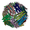 9bpjC  9bpkC 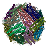 9bq5C C: citing same article ( M: map data used to model this data |
|---|---|
| Similar structure data |


-Links
 PDBj
PDBj


- Assembly
| Deposited unit | 
|
|---|---|
| 1 |
|
-Components
| #1: Protein | Mass: 19917.486 Da / Num. of mol.: 24 Source method: isolated from a genetically manipulated source Source: (gene. exp.) Homo sapiens (human) / Gene: FTL / Production host:  Has protein modification | N | |
|---|
-Experimental details
-Experiment
| Experiment | Method: ELECTRON MICROSCOPY |
|---|---|
| EM experiment | Aggregation state: PARTICLE / 3D reconstruction method: single particle reconstruction |
- Sample preparation
| Component | Name: Human Light Chain Ferritin / Type: COMPLEX / Entity ID: all / Source: RECOMBINANT |
|---|---|
| Molecular weight | Value: .474 MDa / Experimental value: NO |
| Source (natural) | Organism: Homo sapiens (human) |
| Source (recombinant) | Organism: |
| Buffer solution | pH: 7.5 |
| Specimen | Embedding applied: NO / Shadowing applied: NO / Staining applied: NO / Vitrification applied: YES |
| Vitrification | Cryogen name: ETHANE |
- Electron microscopy imaging
| Experimental equipment |  Model: Titan Krios / Image courtesy: FEI Company |
|---|---|
| Microscopy | Model: FEI TITAN KRIOS |
| Electron gun | Electron source:  FIELD EMISSION GUN / Accelerating voltage: 300 kV / Illumination mode: FLOOD BEAM FIELD EMISSION GUN / Accelerating voltage: 300 kV / Illumination mode: FLOOD BEAM |
| Electron lens | Mode: BRIGHT FIELD / Nominal defocus max: 2800 nm / Nominal defocus min: 100 nm |
| Image recording | Electron dose: 9.58 e/Å2 / Film or detector model: GATAN K2 SUMMIT (4k x 4k) |
- Processing
| CTF correction | Type: PHASE FLIPPING AND AMPLITUDE CORRECTION |
|---|---|
| 3D reconstruction | Resolution: 3.3 Å / Resolution method: FSC 0.143 CUT-OFF / Num. of particles: 85039 / Symmetry type: POINT |
